As a mechanical engineer with a background in fluid mechanics, the topic of fluid accumulation and resolution in wet macular degeneration is of particular interest to me. Here, I'll present three cases. The first is fairly typical; the second notes inactive, but growing, lesions; and the final case looks at persistent fluid.
CASE 1
An 88-year-old male, former smoker, presented with 3 weeks of vision loss with a visual acuity of 20/200 OD. He had classic choroidal neovascularization (CNV), with well-demarcated areas of intense hyperfluorescence appearing early, followed by progressive leakage on fluorescein angiography (FA).
He was started on ranibizumab (Lucentis, Genentech), but his vision worsened after the first injection, which I occasionally see. Sometimes, the acuity drops initially, even though the retina becomes dry. At 4-, then 6-week intervals, his acuity recovered stable at 20/50, but extending to 8, or even 7 weeks, he developed subretinal fluid again. He maintains stability on a 6-week regimen.
CASE 2
This was a 77-year-old Caucasian female, nonsmoker, with a reduction in acuity to 20/50 OD, over the course of a week, with a growing pigment epithelial detachment (PED). She had drusen and some diffuse edema, with intraretinal and subretinal fluid. FA showed a diffuse, late-staining, fibrovascular pattern.
With aflibercept (Eylea, Regeneron), she improved a line per month to 20/30. When extended to 6 weeks, the PED grew, with the reappearance of a small amount of subretinal fluid. The acuity remained at 20/30. The PED continued to expand, even at 4-week treatment intervals.
This raises the question: Do you treat the persistent fluid, or could a little bit be protective against atrophy? The jury is still out.
CASE 3
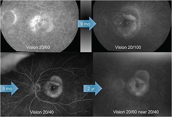
A 78-year-old female, two-pack-per-day smoker was on AREDS2 vitamins for the last year. She presented with multiple large drusen and 20/40 vision in the left eye during her initial visit. Three months later, she developed sudden onset metamorphopsia, noted on a home Amsler grid and 20/60 vision. Late FA showed the combination of a classic appearance CNV and a diffuse multilobed fibrovascular PED.
After a single dose of aflibercept, her visual acuity improved to 20/40, but her PED increased in size; this continued even with 3-week intervals of anti-VEGF. Unfortunately, her acuity continued to decline to 20/100. On FA, her macula developed a blooming rose appearance.
As an alternate approach to treatment, topical difluprednate (Durezol, Novartis) was administered, improving the PED. A month later, she started to decline again, and received an off-label dexamethasone (Ozurdex, Allergan) implant. Again, the PED resolved, with some smoldering, underlying subretinal fluid remaining. Aflibercept at 3-week intervals and dexamethasone implants every 3 months were continued, but neovascularization with late staining surrounding the PEDs persisted. In an effort to treat the PED, photodynamic therapy with a single, half-fluence, 2300-micron spot was administered, with intravitreal triamcinolone afterward to quell the inflammatory response. Two days later there was a hiccup—extensive inflammation and counting fingers vision. Her vision has since recovered to 20/40, and 2 months later she continues to improve (Figure 1). Of note, she quit smoking.

CONCLUSION
When treating AMD, extend treatment to a point but be prepared to treat as frequently as needed. And, be creative.


