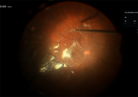
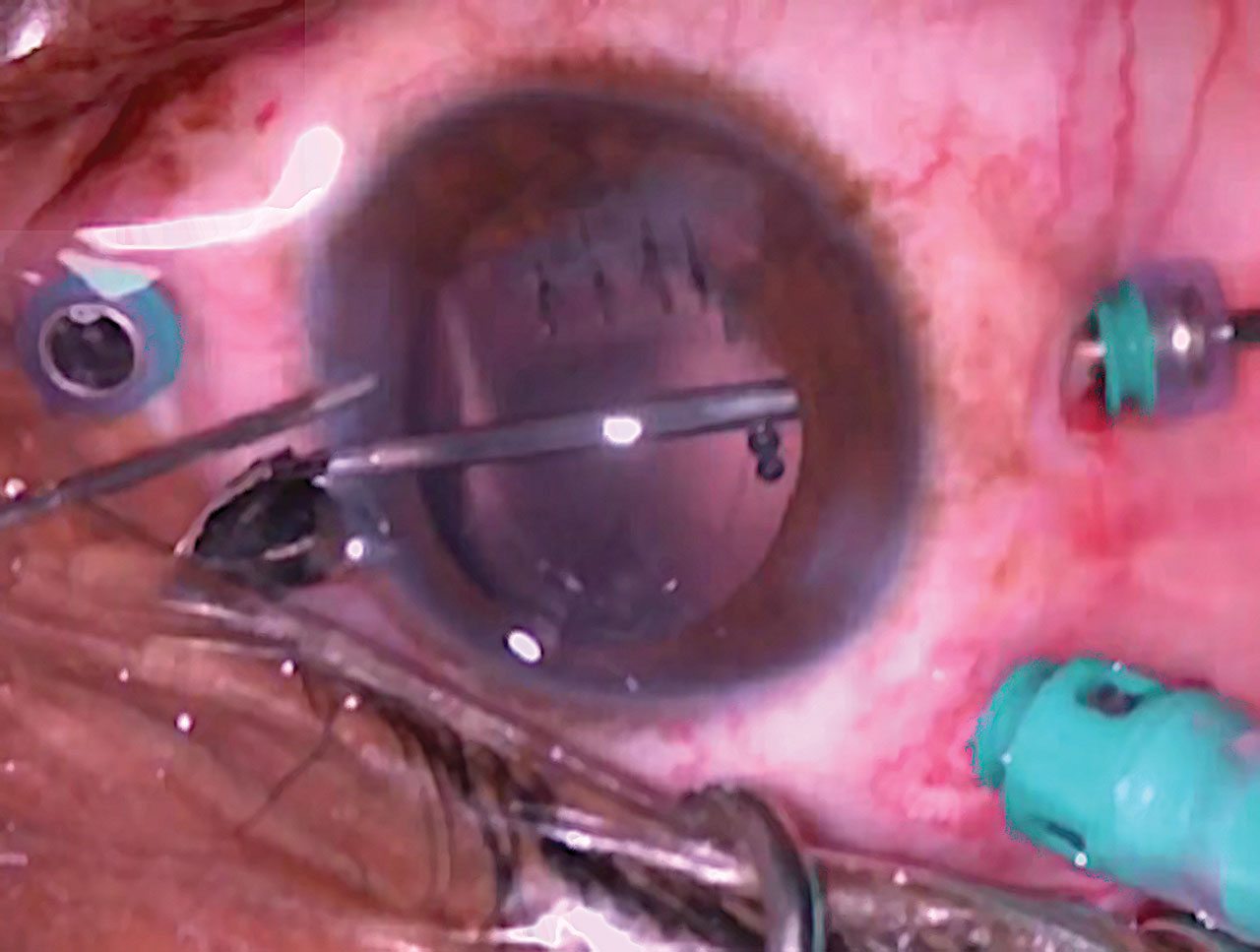
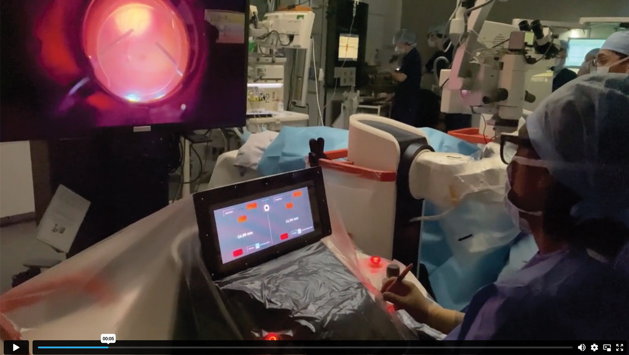
For Retinal Physician's Surgical Pearls video series, Jonathan B. Lin, MD, PhD, demonstrates the proper technique for administering a sub-Tenon’s block, which provides local analgesia and ocular akinesia and is an excellent option for local anesthesia for vitreoretinal surgery, often in conjunction with monitored anesthesia care. Transcript of the narration follows below:
My name is Jonathan Lin, and I am a first-year vitreoretinal surgery fellow at Stanford University. This video demonstrates the proper technique for administering a sub-Tenon’s block. A sub-Tenon’s block provides local anesthesia for vitreoretinal surgery and is preferred by some surgeons as an alternative to a retrobulbar block due to lower risk of globe perforation and orbital hemorrhage, especially for patients on blood thinners. The Tenon’s capsule is a thin layer of connective tissue beneath the conjunctiva that surrounds the globe, and the sub-Tenon’s space is a potential space between the Tenon’s capsule and the sclera. Local anesthetic administered into this area diffuses posteriorly into the retroorbital space and blocks traversing sensory and motor nerves, producing both analgesia and akinesia.
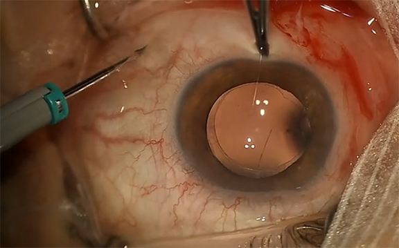
To perform a sub-Tenon’s block, the patient is placed in prone position, and the eye is prepared for surgery in the standard fashion with antiseptic solution followed by placement of a sterile drape. In addition to topical anesthetic eyedrops, we also apply a cotton tip soaked in tetracaine to the planned location of the conjunctival cutdown for additional analgesia. Once analgesia is achieved, a conjunctival cutdown is performed inferonasally. The conjunctiva is tented up with a pair of 0.12 toothed forceps, and a small cutdown is made using blunt Westcott scissors. Gentle blunt dissection is performed until bare sclera is exposed. This patient was a young adult and therefore had relatively robust Tenon’s capsule, which made the dissection more challenging. Once bare sclera has been exposed, the local anesthetic can be applied. We typically use an equal parts mixture of preservative-free 4% lidocaine and 0.75% bupivacaine in a 5 ml syringe with a blunt sub-Tenon’s cannula.
When applying the local anesthetic, little resistance should be encountered, and the anesthetic solution should disappear behind the globe with slight proptosis. If there is resistance, the cannula should be repositioned. If there is ballooning of the conjunctiva, the cannula is in the subconjunctival space as opposed to the sub-Tenon’s space and needs to be repositioned. In some cases, additional cutdown is necessary to reach the appropriate tissue plane. The surgery can now begin. RP


