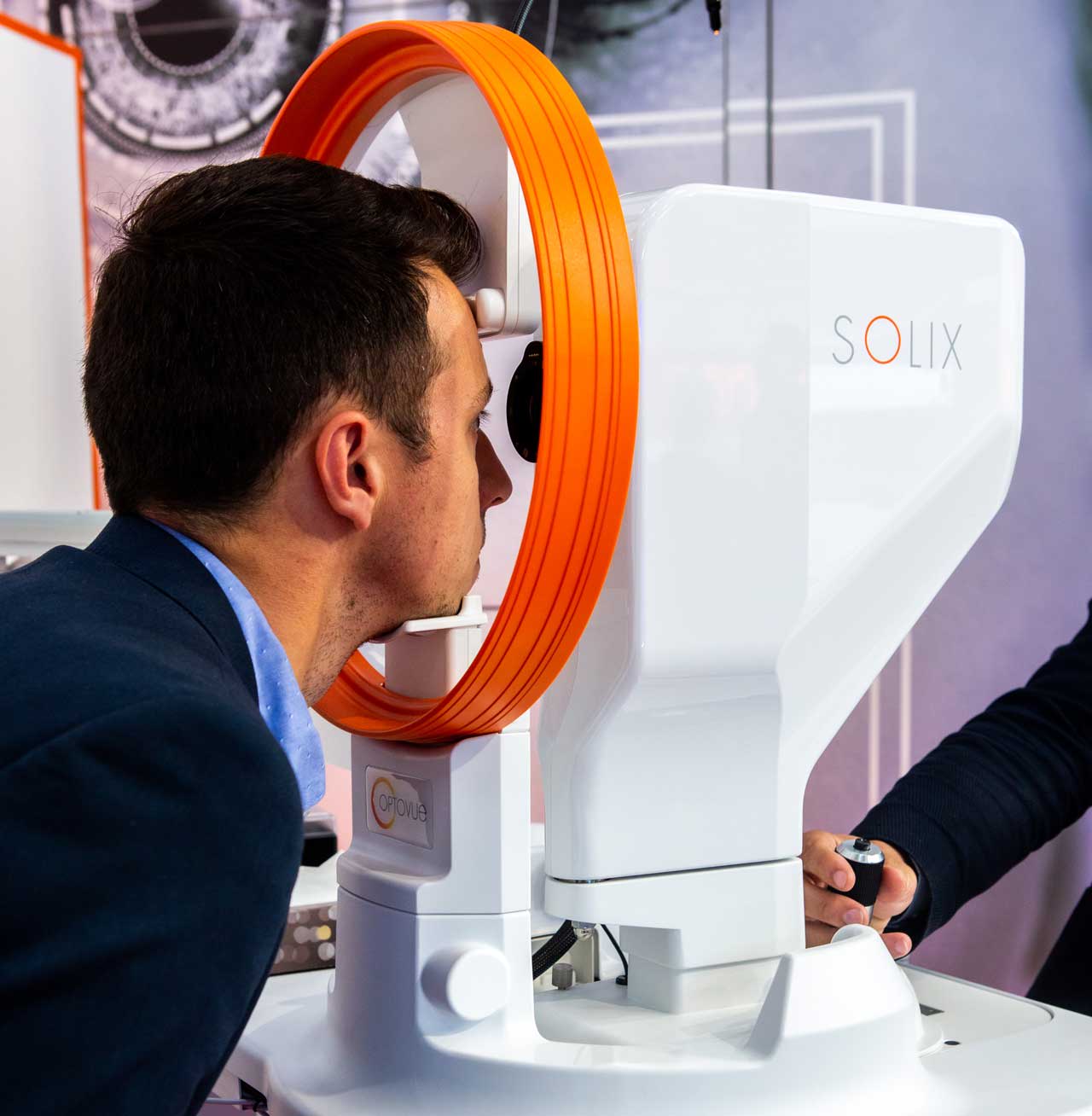
Visionix’s Optovue Solix FullRange optical coherence tomography (OCT) was designed as a next-generation multimodal spectral-domain OCT system to raise the bar in accuracy and efficiency. The system can help detect diseases early as well as monitor and assess advanced ocular pathologies in both the anterior and posterior chambers.

The system provides wide and deep anterior scans (18 mm x 6.25 mm) and posterior scans (16 mm x 6.25 mm), posterior OCT angiography (OCTA), color fundus, external color, and external infrared (IR) imaging. OCTA scans provide structural imaging with an FDA-approved reference database comparison and OCTA vessel density metrics from the same scan. The color camera can capture fundus, disc with overlay imaging, and external color images for documentation, says Peter Naismith, OCT global product manager at Visionix.
“FullRange imaging, coupled with advancements in camera speed (120,000 A-scans per second), allows the Optovue Solix to have larger, denser, more advanced 3D scans with better repeatability and reproducibility compared to its predecessor,” Naismith says.
The Optovue Solix also includes Visionix’s proprietary scans and software — AngioVue, iWellness, and AngioWellness. AngioVue, the only FDA-cleared OCTA metrics, can provide noninvasive 3D visualization and quantification of retinal vasculature. This allows users to assess and confidently manage retinal disease. iWellness and AngioWellness screening software combined with FDA-cleared foveal avascular zone and vessel density metrics provide more advanced 3D analysis of the retinal and optic nerve structures and function to further advance diagnosis with precise imaging.
Features
The Optovue Solix takes its roots from Optovue’s Avanti, the first OCTA system to ever exist, and the only other OCTA with FDA-approved metrics. “The jump in speed from 70,000 A-scans per second to 120,000 A-scans per second offered the opportunity to design new scan patterns at a faster rate,” Naismith says.
Jay Bansal, MD, an ophthalmologist at LaserVue Eye Center in San Francisco, California, who uses the Optovue Solix, says, “It has certainly met my expectations of higher quality resolution and better diagnostics, and it’s easier for my staff to capture the diagnostics.”
Ease of Use
Style and ergonomics were built into the Optovue Solix, such as handholds where patients can reach them. This makes it easier for patients to sit comfortably with a slightly forward position, keeping them securely in the headrest. The keyboard swings in and out for storage but doesn’t need to be moved between scans or patients. The monitor has a wide range of motion and during capture displays both exterior and interior views of the eye with the addition of an easy-to-read working distance and alignment indicators.
Programmable scan protocols and multiple scan features make it easy for technicians to obtain all the necessary images and data for a comprehensive analysis. This can optomize efficiency and streamline exams because less time is needed to move patients to different machines. The system also provides better data than previous versions, and its all-in-one customizable printout enhances efficiency and interpretation of reports.
“Patients are impressed with the level of resolution and the data that we’re able to acquire about their eye,” Dr. Bansal says. “We can re-evaluate the optic nerve or the macula 1 year later and show it to patients and compare the data.”
Clinical Applications
The Optovue Solix can assist in identifying normal results to advanced pathology for both anterior and posterior ocular structures and compares posterior values to an FDA-cleared normative database. It can distinguish structural changes related to retinal pathologies such as diabetic eye diease, schisis, CSR, CME, and wet and dry AMD. RP


