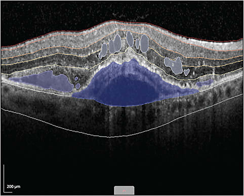
Bern, Switzerland, and Boston-based RetinAI Medical AG (RetinAI) created the software platform Discovery Core to enable clinicians to perform data analysis at an expert level more quickly and efficiently without losing the quality of analyzing disease biomarkers and endpoints, says Nataša Jovic, BSc, MBA, vice president of marketing at RetinAI Medical AG.
“Reading and annotating images to quantify disease endpoints currently requires a lot of time across optical coherence tomography (OCT) scans or per OCT volume; that time can be significant if large data sets need to be reviewed,” Jovic says. Furthermore, “It can be cost prohibitive for most physicians to routinely access services to annotate data to learn more about patient population subgroups based on volumetric measures of biomarkers.”

Discovery Core was also created to overcome challenges related to collaborating with other physicians. Challenges occur, in part, due to the different locations where data reside and how data present in different formats and on different devices. “These obstacles can limit the robustness of insights and knowledge that physicians can gather,” Jovic says.
CLINICAL APPLICATIONS
Discovery Core can be used to design clinical and research studies, compare clinical and real-world evidence, set best practices in clinics for evaluating patient cases, and facilitate teleophthalmology practice to remotely collaborate on data sets in real time across physician groups. “Core can improve patient care by using disease insights that can be generated and shared at a more accelerated pace compared to today’s manual or cumbersome ways for transferring and gaining knowledge,” Jovic says.
Specifically, the platform can be used to monitor patients with macular diseases, such as macular edema, age-related macular degeneration (AMD), geographic atrophy, neovascular AMD, diabetic retinopathy, diabetic macular edema, retinal vein occlusions, MacTel, and other retinal conditions, as well as patients with glaucoma and neuro-ophthalmic diseases. “Disease progression and outcomes can be evaluated over time to better understand patient subgroups based on biomarkers and endpoints,” says Marion R. Munk, MD, PhD, a medical retina and uveitis specialist at Augenarzt-Praxisgemeinschaft Gutblick AG in Pfäffikon, Switzerland, who helped to validate the Core platform.
Artificial intelligence (AI) models for fluid and layer segmentation automatically enrich OCT images across B-scans and provide volumetric measures of subretinal fluid, intraretinal fluid, pigment epithelium detachment, and thickness measures for 5 layers of the retina. Physicians can review progression plots of these biomarkers and endpoints across visits to analyze outcomes over time, Jovic says. Additional clinical information can be added through electronic case report forms that can be customized and attached to each patient visit. Beyond AI, users can use a set of annotation tools to segment biomarkers on imaging data sets, which provides a means to develop new AI models.
HOW IT WORKS
Using AI, the Discovery Core provides an automated and immediate analysis of fluid and layer segmentation of OCT volumes in the retina. The cloud-based platform provides real-time collaboration across physicians’ data sets, Jovic says. Large-scale analysis is immediately accessible for in-depth, biomarker, and endpoint measures on patient subgroups in a secure software environment, which can be shared to help advance patient management.
According to Dr. Munk, the software platform is easy to use. Users can upload images as well as OCT and fundus images via a drag-and-drop feature. Optical coherence tomography images are processed right away and are analyzed with AI. “Physicians have an almost instantaneous output to layer thicknesses and fluid volume measures,” she says.
If images of multiple visits are uploaded, users can easily compare individual visits with each other and visualize the volumetric data of layer thicknesses and fluid volumes plotted over time, which gives immediate insight on a patient’s current and previous disease activity, Dr. Munk continues. This facilitates treatment decisions and treatment planning, as well as design of new areas of academic and clinical research.
Immediate image and data sharing is done securely and follows HIPAA regulations. “Multiple users can work simultaneously on a large, shared imaging data set,” Dr. Munk says.
Regarding clinical trials, the Core platform allows physicians to instantly access uploaded images in real time while a trial is still ongoing. “Physicians can assess a drug’s safety and evaluate immediate indicators of efficacy to better understand how a new drug is potentially working,” Dr. Munk says. “The software allows physicians to reassess a study’s procedures and enables quick adjustments in terms of study amendments if necessary.”
EASE OF USE
Discovery Core has an intuitive user interface that can be easily navigated, organizing data into study workbooks or folders with review screens that provide side-by-side comparisons of imaging data, Jovic says. These comparative views can simultaneously be evaluated across B-scans so users are at the same location of the scan throughout the analysis.
Progression plots of biomarkers can be viewed in a single instance, and when a particular visit is clicked on the plot, a more detailed view of the image and visit can be examined, Jovic says. Grading surveys can be deployed across reviewers, and the status of a review can be monitored. Core was designed to be user friendly, with analysis tools easily accessible and data and insights shared in an immediate and frictionless manner.
With the Core platform, Dr. Munk says she can include more sites and clinics for multicenter studies, because various OCT devices can be used in one study and reviewed simultaneously. “It’s no longer necessary to have software for multiple OCT devices, because all types of images can be processed on Core, providing immediate access to all images for all study participants,” she says.
WHAT STUDIES SHOW
Discovery Core’s AI models have been studied1 in multiple diseases of the retina, including macular edema, wet AMD, geographic atrophy, diabetic retinopathy, and retinal vein occlusions. Artificial intelligence models achieved measures that were similar to expert human graders with more than 95% agreement in most cases of evaluating disease biomarkers and endpoints.2
“AI models provide the ability for physician researchers to consistently and in a standardized way evaluate large data sets without fatigue to help advance our understanding of disease,” Jovic concludes. RP
REFERENCES
- Apostolopoulos S, Salas J, Ordóñez JLP, et al. Automatically enhanced OCT Scans of the retina: a proof of concept study. Sci Rep. 2020;10(1):7819. doi:10.1038/s41598-020-64724-8
- Mantel I, Mosinska A, Bergin C, et al. Automated quantification of pathological fluids in neovascular age-related macular degeneration, and its repeatability using deep learning. Transl Vis Sci Technol. 2021;10(4):17. doi:10.1167/tvst.10.4.17


