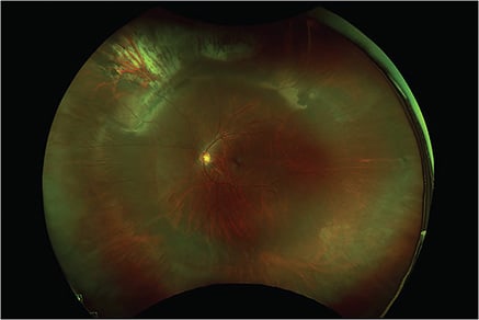
Scleral buckling (SB) surgery is an important surgical tool to fix rhegmatogenous retinal detachments (RRD). Scleral buckling surgery can be used in combination with pars plana vitrectomy (PPV) or as a standalone surgery (primary SB). Primary SB surgery has a high single-surgery success rate.1 However, data show that a majority of surgeons are placing a SB in less than 20% of RRDs.2,3
Due to the decline in primary SB surgery, graduating fellows might see less SB surgery, which translates to being less prepared to anticipate and manage potential complications. This article provides surgical pearls for a few common and rare SB complications.
FINDING AND TREATING THE BREAK
Preoperative planning is essential in all cases of retinal detachments. Identifying the retinal break in clinic allows for appropriate surgical planning and decreases risk of redetachment. Intraoperatively, proper SB placement depends on identifying, marking, and treating the retinal break. It is routinely done with indirect ophthalmoscope.
Recently, several reports have advocated for visualizing and treating the break using a noncontact wide-angle viewing system with a cannula-based chandelier endoilluminator.4 When treating a break with cryotherapy, one should be aware of the position of the probe tip. Mistaking the shaft of the probe for the probe tip could result in posterior cryotherapy scars including in the macular region.5
CONJUNCTIVA SCARRING AND GLAUCOMA FILTRATION SURGERY
Assessment of the conjunctiva and sclera should be done preoperatively with the use of the slit lamp. Any prior glaucoma filtration surgery, strabismus surgery, or extensive conjunctival scarring should be noted. Retinal detachments occurring after glaucoma tube shunt implantation require appropriate preoperative counseling, given the lower single-surgery success rates and poor visual outcomes in these patients.6-8 Care should be taken to avoid disturbing the area of prior surgery and, in most cases, this involves performing a PPV with careful maneuvering around the tube shunt or trabeculectomy site for proper cannula placement. If a scleral buckle is to be performed, circumferential or radial segmental elements can be used to support retinal breaks or pathology in quadrants other than the implant device. Meticulous attention to the conjunctiva is necessary if conjunctival peritomy is to be performed with a tube shunt in place to avoid exposure and manipulation of the encapsulated bleb. If the surgeon feels that 360-degree SB support is needed, the tube shunt occasionally may need to be removed to make room for the SB.

CUT RECTUS MUSCLE
Starting the conjunctival peritomy at the limbus minimizes the risk of inadvertently cutting a rectus muscle. Prior strabismus surgery history should be elicited. Gentle muscle identification and isolation can prevent splitting of the muscles and potential diplopia. However, in rare cases of cut rectus muscle, if the proximal end has not been lost, the muscle can be imbricated with a Vicryl suture and reanchored.
If the proximal end of the muscle is lost, careful exploration with a hand-over-hand technique should be performed. The microscope can be helpful, especially to avoid excessive manipulation of Tenon’s capsule to reduce the risk of developing fat adherence syndrome. If the muscle is retrieved, it can be reattached to the globe. If the muscle is lost, the surgeon should consider intraoperative assistance from a strabismus surgeon or an expedited evaluation. If the muscle cannot be retrieved and the patient has symptomatic diplopia, elective transposition surgery can often be performed.9
THIN SCLERA
In pronounced cases of scleral thinning, the purplish hue of the choroid can be appreciated on clinical preoperative exam. Patients with connective tissues disorders, high myopes, and patients with prior scleral surgery are at particular risk. Intraoperatively, sclera should be examined after the rectus muscles are isolated. It could be done with a Schepens orbital retractor or cotton tip applicators. In most cases with scleral ectasia, the suture pass or belt loop may be repositioned away from an area of concern. Alternatively, a segmental element may be utilized instead of the circumferential buckle if the breaks are localized in an area without scleral thinning. Sometimes, if a quadrant has severe thinning, that quadrant can be left unsutured, especially if the retinal breaks are not in this quadrant. This, however, can result in anterior migration of the buckle and potentially lesser buckle effect. For surgeons who use belt loops, significant thinning may lead them to avoid belt loops and use sutures.
Regarding suturing, the use of a spatulated needle with its side-cutting design is very important to best facilitate a clean partial-thickness pass through the sclera. When passing the needle through the sclera, it is imperative to make sure the heel of the needle does not penetrate the sclera by following the curve of the needle as it is pulled through. One should avoid placing a suture near the vortex veins because of the risk of suprachoroidal hemorrhage. In very rare instances of significant thinning (such as in a scleromalacia patient), the buckle portion may need to be aborted and the case may be converted to a primary PPV. It is important to remember that particularly in cases of thin sclera, judicious cryotherapy and sufficient time (and balanced salt solution [BSS] application) allowing for probe thawing are key to avoid scleral perforation. Trying to remove the probe prematurely can also result in the choroidal vessel rupture and subretinal hemorrhage.
DEEP SCLERAL SUTURE PASS WITH AND WITHOUT SUBRETINAL BLEEDING
In the event of a deep suture pass, intraocular pressure (IOP) control should be the first concern, and the following steps should be performed in an expedient but safe manner. The suture should be removed and pressure should be maintained by applying a cotton tip to the site of perforation while pulling on the silk sutures used to isolate the rectus muscles. Preventing hypotony is important to avoid new or worsening subretinal and/or choroidal hemorrhage. If the above methods cannot control the IOP, sterile BSS should be injected via the pars plana to repressurize the globe. Occasionally, the perforation site might need to be sutured. Upon reestablishing the IOP, an indirect ophthalmoscopy should be performed to examine for iatrogenic breaks and subretinal or submacular hemorrhage. If an iatrogenic break at the site of a deep pass is visualized, then cryotherapy should be applied. Frequently, extramacular bleeds can be observed. In cases of small and thin submacular bleed, gas tamponade can be used with face-down positioning to aid in blood displacement. If the hemorrhage appears significant, a PPV can be considered, and drainage through a posterior retinotomy has been described.10 Once attention is returned to passing the remaining sutures, ensuring proper positioning of the buckle over the area of the deep pass is important.
RETINAL INCARCERATION
The highest risk for retinal incarceration occurs during sub-retinal fluid drainage, which is usually performed with cut-down or needle-drainage techniques. The former tends to have a higher risk of incarceration. When incarceration occurs, it is important to support the affected area with the scleral buckle and to treat it with cryotherapy; this may require repositioning, although ideally the drainage site was already supported by the buckle. If the incarcerated area is significant, various techniques have been described in isolated cases to dislodge the retina, including external injection of viscoelastic into the perforation site or positive fluid pressure through the sclerotomy.10,11 In most cases, small incarcerations will settle in the postoperative period with appropriate support. In persistent cases, PPV can be performed with a localized retinotomy over the incarcerated site to flatten the retina.10
CONCLUSION
Scleral buckling will continue to be a vital tool in the armamentarium of the modern-day retinal surgeon. Some RRD presentations might benefit from primary scleral buckle surgery or combined buckle and PPV. Surgeons’ ability to anticipate and manage potential complications will ensure best outcomes for patients requiring SB surgery. RP
REFERENCES
- Ryan EH, Joseph DP, Ryan CM, et al. Primary retinal detachment outcomes study: methodology and overall outcomes-primary retinal detachment outcomes study report number 1. Ophthalmol Retina. 2020;4(8):814-822. doi:10.1016/j.oret.2020.02.014
- McLaughlin MD, Hwang JC. Trends in vitreoretinal procedures for medicare beneficiaries, 2000 to 2014. Ophthalmology. 2017;124(5):667-673. doi:10.1016/j.ophtha.2017.01.001
- Singh RP, Stone TW, eds. ASRS 2018 preferences and trends membership survey. Chicago: American Society of Retina Specialists; 2018.
- Caporossi T, Finocchio L, Barca F, Franco F, Tartaro R, Rizzo S. Scleral buckling for primary rhegmatogenous retinal detachment using a noncontact wide-angle viewing system with a cannula-based 27-g chandelier endoilluminator. Retina. 2019;39 Suppl 1:S144-S150. doi:10.1097/IAE.0000000000001891
- Boscia F, Giacipoli E, Ricci GD, Sborgia G. Management of intraoperative complications during scleral buckling surgery. In: Patelli F, Rizzo S, eds. Management of Complicated Vitreoretinal Diseases. Springer International Publishing; 2015:103-109. Accessed February 11, 2022. http://link.springer.com/10.1007/978-3-319-17208-8_8
- Waterhouse WJ, Lloyd MA, Dugel PU, et al. Rhegmatogenous retinal detachment after Molteno glaucoma implant surgery. Ophthalmology. 1994;101(4):665-671. doi:10.1016/s0161-6420(94)31280-2
- Benz MS, Scott IU, Flynn HW Jr, Gedde SJ. Retinal detachment in patients with a preexisting glaucoma drainage device: anatomic, visual acuity, and intraocular pressure outcomes. Retina. 2002;22(3):283-287. doi:10.1097/00006982-200206000-00005
- Sharma U, Panda S, Balekudaru S, Lingam V, Bhende P, Sen P. Outcomes of rhegmatogenous retinal detachment surgery in eyes with pre-existing glaucoma drainage devices. Indian J Ophthalmol. 2018;66(12):1820-1824. doi:10.4103/ijo.IJO_438_18
- Ellis EM, Kinori M, Robbins SL, Granet DB. Pulled-in-two syndrome: a multicenter survey of risk factors, management and outcomes. J AAPOS. 2016;20(5):387-391. doi:10.1016/j.jaapos.2016.06.004
- Najafi M, Mammo DA, Emerson GG. Management of suture penetration in combined vitrectomy and scleral buckle surgery. J Vitreoret Dis. 2020;4:202-209.
- Stopa M, Toth CA. A method to free retina and vitreous from intraoperative incarceration in the sclerotomy. Retina. 2006;26(9):1070-1071. doi:10.1097/01.iae.0000247195.28648.eb


