Since the introduction of the first 27-gauge vitrectomy system in 2010, significant advances have been made in instrumentation and surgical techniques using this approach. Initially, 27-gauge vitrectomy was used only for select cases, such as macular pathologies and noncomplex vitreous hemorrhage.1 Early reports focused on the “feasibility” of 27-gauge vitrectomy. However, as its efficacy, safety profile, and favorable visual and anatomic outcomes were realized, use of 27-gauge vitrectomy expanded to more challenging cases, including diabetic traction retinal detachment (TRD) and primary rhegmatogenous retinal detachment with or without proliferative vitreoretinopathy (PVR).2,3
The development of high-speed cutters and a wide variety of other 27-gauge instruments, in conjunction with new vitrectomy machines, helped this transition. Long-term follow-up data of large cohorts became available, which demonstrated the long-term efficacy and safety of 27-gauge systems when used to treat various vitreoretinal diseases.4 In addition, as surgeon experience with 27-gauge vitrectomy increased, applications of its use further expanded to include 27-gauge cannula-assisted intraocular lens fixation, intraocular tumor biopsy using the 27-gauge vitreous cutter, and hybrid surgery using the 27-gauge cutter through 23-gauge or 25-gauge cannulas.5-8 However, extensive cases such as open-globe injury and PVR requiring silicone oil tamponade are likely still not ideal for 27-gauge platforms. This article will review the background, recent advances, and current state of 27-gauge vitrectomy and discuss the specifications of current 27-gauge vitrectomy systems.
THE GENESIS OF 27-GAUGE VITRECTOMY
The driving force behind the initial development of a 27-gauge vitrectomy system by Oshima and associates was not simply the pursuit of smaller incisions, but also the desire to increase the safety, efficacy, and quality of transconjunctival sutureless vitrectomy systems.1 The goal was to reduce the risk of wound sealing–related complications such as hypotony, choroidal detachment, and endophthalmitis, even after extensive vitrectomy and without suturing, while achieving favorable and early visual recovery. The primary obstacle was reduced endoillumination due to the limited number of optic fibers that could fit inside the smaller-diameter light pipes. However, the development of more powerful illumination tools that utilized xenon light (including the Accurus High Brightness Illuminator from Alcon, the Photon from Synergetics, and the Brightstar from DORC), along with the 27-gauge chandelier probe introduced in the mid-to-late 2000s, provided the impetus to continue refining and improving 27-gauge vitrectomy systems.
Early concerns over 27-gauge vitrectomy included instrument fragility and relatively low efficiency of the early-generation cutters. However, the development of high-speed cutters, high-performance vitrectomy machines, and stiffer instruments have significantly improved the efficiency of vitrectomy, enabling surgeons to perform 27-gauge vitrectomy for a wide variety of vitreoretinal diseases.
RECENT ADVANCES IN 27-GAUGE VITRECTOMY SYSTEMS
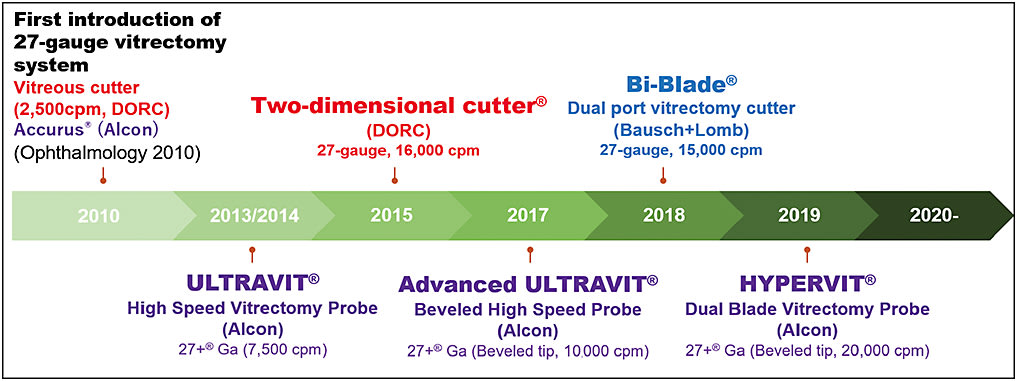
The first-generation 27-gauge system, introduced in 2010, used the Accurus vitrectomy machine and DORC vitreous cutter with a maximum cut rate of 2,500 cuts per minute (cpm).1 The current generation of machines provide significantly higher cutting speeds and improved fluidics; these platforms include the Constellation Vision System (Alcon), Eva (DORC), and Stellaris PC (Bausch + Lomb). Vitreous cutters have continuously improved over time (Figure 1), and the cutting speeds have increased from 2,500 cpm to 10,000 cpm with the single-blade cutter. Furthermore, dual-cutter technology provides a higher cutting speed, high duty cycle, and reduced retinal traction, thereby allowing safe and efficient dissection of the vitreous and membranes even over mobile detached retina. Currently, the 2-dimensional cutter (16,000 cpm) from DORC, the Hypervit dual-blade vitrectomy probe (20,000 cpm) from Alcon, and the Bi-Blade dual-port vitrectomy cutter (15,000 cpm) from Bausch + Lomb are commercially available. Another advance in the 27-gauge vitreous cutter design came with Alcon’s introduction of beveled tips, which enable the cutter port to get closer to the retina. This facilitates access to tighter tissue planes, which is particularly useful in TRD dissections.

In addition to the vitrectomy machine and the vitreous cutter, a variety of 27-gauge instruments, including valved cannulas, various membrane forceps, curved laser probes, diathermy probes, curved scissors, soft-tipped cannulas, light pipes, and various chandeliers have become commercially available from multiple manufacturers. Long-shaft forceps are also available for highly myopic patients. All these instruments are available for 27-gauge vitrectomy, so surgeons can now select the optimal combination of instruments for each case.
CURRENT STATE OF 27-GAUGE VITRECTOMY
Advances in high-speed cutters and instrumentation have negated many of the limitations of the initial 27-gauge vitrectomy system. Many surgeons are now experiencing a noticeable increase in vitreous aspiration flow with current cutters. Looking at Alcon’s Constellation cutters (Figure 2), a recent study reported that the 27-gauge Hypervit dual-blade vitrectomy probe (20,000 cpm) provides a 26% greater maximum flowrate compared to the single-blade 27-gauge Advanced Ultravit beveled high-speed probe (10,000 cpm) and a 48% greater maximum flowrate than the Ultravit probe (7,500 cpm). The 27-gauge Hypervit probe was approximately 83% as efficient as a 25-gauge Advanced Ultravit probe at 10,000 cpm. In addition, there was a 31% reduction in peak traction force for the 27-gauge Hypervit probe compared to the Advanced Ultravit probe in core vitrectomy mode.9

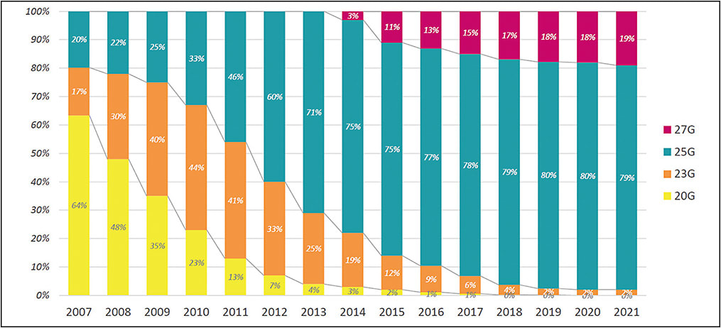
Surgeons have also learned that placing the trocars closer to the 3 o’clock and 9 o’clock meridians to maximize the instrument range and using chandelier endoillumination with wide-angle viewing systems while limiting movement of the eye can facilitate vitreous shaving and intraocular procedures with 27-gauge instruments while minimizing issues related to any instrument flexibility. Consequently, more surgeons are now adopting the 27-gauge system. In Japan, approximately 19% of vitrectomies are now performed with 27-gauge platforms (Figure 3).

Long-term safety profiles for 27-gauge vitrectomy are now available also.4 Overall, the rate of postoperative complications is low across various surgical indications, including macular diseases, diabetic TRD, and complex retinal detachments. Although there is limited evidence to support that 27-gauge surgery has fewer intraoperative complications compared to 23-gauge or 25-gauge vitrectomy, only a few postoperative complications have been reported, including transient ocular hypertension, vitreous hemorrhage, transient hypotony, and retinal redetachment, all of which are expected in a limited fashion. These complications are not specifically related to the use of 27-gauge systems.4,10
The benefit of 27-gauge vitrectomy in preventing postoperative hypotony has been evaluated in many studies. A recent prospective randomized trial confirmed that 27-gauge vitrectomy is associated with smaller reduction in postoperative intraocular pressure (IOP).11 The mean changes in pressure before and immediately after surgery in the 27-gauge (n=67) and 23-gauge (n=68) groups were -0.40 mmHg and -3.05 mmHg, respectively (P=.013). A few patients in the 23-gauge group, but none in the 27-gauge group, required suturing of their sclerotomies. The reduced incidence of postoperative hypotony after 27-gauge vitrectomy is theoretically expected to decrease the incidence of postoperative endophthalmitis. Although no studies have yet evaluated the incidence of endophthalmitis after 27-gauge vitrectomy, experimental studies with human cadaver eyes have shown that bacterial entry into the eye is less likely with 27-gauge perpendicular (nonbeveled) incisions compared to 23-gauge perpendicular incisions.12
Currently, the indications for 27-gauge vitrectomy comprise almost all vitreoretinal diseases requiring surgery, including macular diseases (epiretinal membrane, macular hole, myopic traction maculopathy), vitreous opacities, vitreous hemorrhage, intraocular lens dislocation, submacular hemorrhage, diabetic TRD, primary rhegmatogenous retinal detachment, and PVR (Figure 4). However, based on our experience, eyes with open-globe injuries with massive subretinal hemorrhage and choroidal hemorrhage and cases of severe PVR that require silicone oil tamponade tend not to be ideal for 27-gauge surgery. Very dense and thick tissues may still be challenging for the 27-gauge cutter to handle, and while silicone oil insertion and removal through 27-gauge cannulas is possible, it takes longer.

27-GAUGE VITRECTOMY FOR CHALLENGING CONDITIONS
Diabetic Traction Retinal Detachment
The small and beveled tip of 27-gauge cutters is an elegant approach for diabetic TRD because it can be easily inserted into the tight spaces between fibrovascular pegs where larger instruments cannot enter.13 The 27-gauge beveled tip is also particularly useful for separating the posterior hyaloid-fibrovascular membrane complex from the retina because the short distance from the cutter port to the retinal surface allows access to the tissue plane without damaging the retina. The recent development of higher cutting rates facilitates the dissection of fibrovascular membranes from the retinal surface, while minimizing the risk of iatrogenic breaks. The multifunctionality of the cutter allows it to serve as a membrane pick, forceps, soft-tip, and scissors, thereby contributing to streamlined surgeries that require few instrument exchanges. The prevention of postoperative hypotony with 27-gauge vitrectomy also has the potential to prevent rebleeding. The preservation of the conjunctiva without scarring is an additional benefit, because some patients may have glaucoma surgery in the long term. Numerous studies have reported the feasibility, safety, and favorable visual outcomes of 27-gauge vitrectomy for diabetic TRD.4,10
Primary Rhegmatogenous Retinal Detachment
The small port, in conjunction with the high-speed cutting rate of a 27-gauge vitrector, is especially useful for efficient shaving of the vitreous over the detached retina, while lowering the risk of iatrogenic breaks because of the reduction in retinal mobility. A recent multicenter study that evaluated the feasibility and safety of 27-gauge vitrectomy in 410 cases of primary rhegmatogenous retinal detachment reported that primary reattachment was achieved in 392 eyes (96%) and final reattachment was achieved in 410 eyes (100%).14 All surgeries were completed using a 27-gauge system without sclerotomy suturing or any need for conversion to a larger-gauge system. The authors of this study used contact or noncontact wide-angle viewing systems in all cases to perform surgery with 27-gauge instruments without tilting the eye. Several other studies have also reported the safety and feasibility of 27-gauge vitrectomy for giant-tear retinal detachment and PVR.4,15 Similar to the benefits provided by 27-gauge cutters for diabetic TRD, a small port close to the tip and high-speed cutting rate provided efficient shaving of vitreous and proliferative membranes from the retinal surface with fewer instrument exchanges.
Other Indications
A 27-gauge vitreous cutter is useful for the biopsy of intraocular tumors, such as uveal melanomas, for molecular prognostication. A recent study reported that a 27-gauge cutter can obtain tissue samples with favorable cellularity and diagnostic adequacy.5
Secondary intraocular lens fixation can also be performed with a Gore-Tex suture (W.L. Gore) and the Yamane technique in the setting of 27-gauge vitrectomy. The 27-gauge cannula-based intraocular lens fixation is another useful technique for patients with dislocated intraocular lenses.7,8
Certain cases of pediatric vitrectomy may be beneficial to perform with 27-gauge platforms also. Complete separation of the hyaloid is challenging with any gauge system in young children, and particularly 27-gauge. Therefore, in older children where the hyaloid can be more easily addressed or cases where hyaloid separation is not required, 27-gauge surgery offers the benefits of reliable sutureless surgery with improved postoperative comfort. An easier postoperative course facilitates better compliance from children who may be reluctant to position or use eye drops if uncomfortable. We recently reported on a case series of pediatric vitrectomies for a wide range of diagnoses, which showed the feasibility and safety of pediatric 27-gauge surgery.16
CONCLUSIONS
The latest advances in 27-gauge vitrectomy have made possible the many benefits intended to be expected of transconjunctival sutureless vitrectomy, such as better sealing sclerotomies, reduced hypotony, less rebleeding after diabetic vitrectomy, fewer iatrogenic breaks, and favorable visual recovery. The evolution of 27-gauge vitrectomy over the last decade has been tremendous, and further developments to achieve even greater fluidics, stiffer instruments, and higher cut rates will continue over the coming years. RP
REFERENCES
- Oshima Y, Wakabayashi T, Sato T, et al. A 27-gauge instrument system for transconjunctival sutureless microincision vitrectomy surgery. Ophthalmology. 2010;117(1):93-102.e2. doi:10.1016/j.ophtha.2009.06.043
- Khan MA, Shahlaee A, Toussaint B, et al. Outcomes of 27 gauge microincision vitrectomy surgery for posterior segment disease. Am J Ophthalmol. 2016;161:36-43.e2. doi:10.1016/j.ajo.2015.09.024
- Yoneda K, Morikawa K, Oshima Y, et al. Surgical outcomes of 27-gauge vitrectomy for a consecutive series of 163 eyes with various vitreous diseases. Retina. 2017;37(11):2130-2137. doi:10.1097/IAE.0000000000001442
- Khan MA, Kuley A, Riemann CD, et al. Long-term visual outcomes and safety profile of 27-gauge pars plana vitrectomy for posterior segment disease. Ophthalmology. 2018;125(3):423-431. doi:10.1016/j.ophtha.2017.09.013
- Klofas LK, Bogan CM, Coogan AC, et al. Instrument gauge and type in uveal melanoma fine needle biopsy: implications for diagnostic yield and molecular prognostication. Am J Ophthalmol. 2021;221:83-90. doi:10.1016/j.ajo.2020.08.014
- Yonekawa Y, Thanos A, Abbey AM, et al. Hybrid 25- and 27-gauge vitrectomy for complex vitreoretinal surgery. Ophthalmic Surg Lasers Imaging Retina. 2016;47(4):352-355. doi:10.3928/23258160-20160324-08
- Jujo T, Kogo J, Sasaki H, et al. 27-gauge trocar-assisted sutureless intraocular lens fixation. Bmc Ophthalmol. 2021;21(1):8. doi:10.1186/s12886-020-01758-6
- Diamint DV, Giambruni JM. 27-gauge trocar-assisted transconjunctival sutureless intraocular lens scleral fixation. Eur J Ophthalmol. 2020;31(3):NP65-NP69. doi:10.1177/1120672120919068
- Inoue M, Koto T, Hirakata A. Comparisons of flow dynamics of dual-blade to single-blade beveled-tip vitreous cutters. Ophthalmic Res. 2022;65(2):216-228. doi:10.1159/000521468
- Cruz-Iñigo YJ, Berrocal MH. Twenty-seven-gauge vitrectomy for combined tractional and rhegmatogenous retinal detachment involving the macula associated with proliferative diabetic retinopathy. Int J Retina Vitreous. 2017;3:38. doi:10.1186/s40942-017-0091-x
- Charles S, Ho AC, Dugel PU, et al. Clinical comparison of 27-gauge and 23-gauge instruments on the outcomes of pars plana vitrectomy surgery for the treatment of vitreoretinal diseases. Curr Opin Ophthalmol. 2020;31(3):185-191. doi:10.1097/ICU.0000000000000659
- Cohen MN, Houston SK 3rd, Roberts AL, et al. Analysis of pars plana vitrectomy incisions using live bacteria. Retina. 2017;37(6):1152-1159. doi:10.1097/IAE.0000000000001319
- Oshima Y. Use of 27-gauge vitrectomy for diabetic TRD. Retina Today. 2015;10(6):33-36.
- Shinkai Y, Oshima Y, Yoneda K, et al. Multicenter survey of sutureless 27-gauge vitrectomy for primary rhegmatogenous retinal detachment: a consecutive series of 410 cases. Graefe’s Archive Clin Exp Ophthalmol. 2019;257(12):2591-2600. doi:10.1007/s00417-019-04448-2
- Kunikata H, Aizawa N, Sato R, Nishiguchi KM, Abe T, Nakazawa T. Successful surgical outcomes after 23-, 25- and 27-gauge vitrectomy without scleral encircling for giant retinal tear. Jpn J Ophthalmol. 2020;64(6):506-515. doi:10.1007/s10384-020-00755-y
- Yonekawa Y, Berrocal AM, Kusaka S, et al. 27-gauge vitrectomy for pediatric vitreoretinopathies. Presented at: Retina Society 2017 annual meeting, Boston, MA.


