An efficient peripheral vitrectomy is a crucial step when performing thorough vitrectomy for many vitreoretinal conditions. Traditionally, this is either done via scleral indentation that is performed by a surgical assistant or by using a chandelier light fiber.
Challenge
Having a skilled surgical assistant or using a chandelier endoillumination system to assist in peripheral vitrectomy is a luxury that many vitreoretinal surgeons do not have in developing countries. This is mainly due to the availability of trained staff and high costs involved with additional instrumentation. In the last few years, independent shaving of peripheral vitreous have become the norm, because various models of light pipe sleeves have come into the market that enable independent peripheral vitrectomy by means of illuminated scleral depressing. Despite this, the cost remains prohibitive for surgeons working in developing countries.
Solution
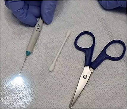
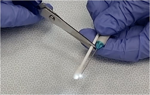
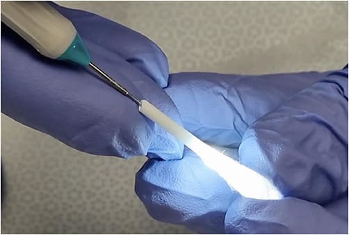
There is a need for an alternative to address this gap. This is where the modified illuminated scleral depressor has a role to play. This modification involves using a sterile cotton tip with a hollow plastic shaft (Figure 1). This is then measured by placing the cotton tip next to the light pipe and subsequently trimmed to the length of the light pipe (Figure 2; measured from tip to the base of the light pipe). The light pipe is then inserted into the shaft (Figure 3). A sterile tape can be used to secure the shaft to the light pipe or alternatively the surgeon can use a finger to stabilize it. The diameter of the cotton tip shaft can accommodate light pipes of all gauges.


Tips for Modified Illuminated Scleral Depressor
Wetting the cotton tip with saline prior to using it on the surface of the eye will improve maneuverability on surface of the conjunctiva. The illumination settings are then increased to the maximum allowable levels by the respective machines. Always remember to reset the illumination levels to baseline before reintroducing the light pipe into the vitreous cavity. Rotating the eye to the opposite side of the area of interest makes it easier to slide the depressor in place before centering the eye.
Based on the authors’ experience, the visualization of the peripheral vitreous and retina with this modification is comparable to commercially available illuminated scleral indenter sleeves (Video 1). Admittedly, visualization of the peripheral vitreous does not compare with endoillumination, but prior staining with triamcinolone allows for better visualization of the vitreous during this maneuver. The authors have not encountered any complications while using this device. However, one of the drawbacks of using a cotton tip is that it tends to soak up blood that may be in the surgical field and this causes the illumination to be dimmed over time. A simple change to another cotton tip will resolve this issue. Using the sleeve without taping it down will need some practice, but the authors have never experienced dislodgement when using a middle finger to provide additional support.
Conclusion
In terms of visualization, this modification is a comparable alternative to the sleeve options currently on the market for independent peripheral vitrectomy, especially in cases where trimming of the vitreous base is required. It is an excellent low-cost option that is a useful addition in the armamentarium of vitreoretinal surgeons, especially in developing countries.


