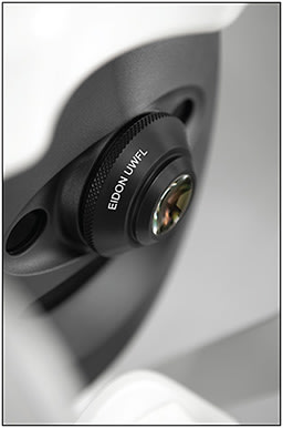
When users of iCare’s Eidon platform asked for a better and easier way to see the far periphery of the retina, the company listened. Ultrawidefield devices on the market can compromise in either image quality or ease of use, says Luca Zalunardo, vice president of marketing for iCare USA in Fremont, California. iCare was able to expand the platform’s retina coverage while retaining its usability and image quality by introducing a module with 2 core innovations: TrueColor confocal imaging and automation in ultrawidefield imaging.
Ultrawidefield imaging can show retinal anatomic features anterior to the vortex vein ampullae in all 4 quadrants. “With its ultrawidefield lens accessory, the Eidon can cover this surface in real color and on undilated patients,” Zalunardo says. “This is particularly important to detect pathologies that affect the peripheral retina, such as diabetic retinopathy and retinal detachments.”
Because of its capabilities, Altin Pani, MD, a retinal physician at Retina Consultants in Providence, Rhode Island, tried the new Eidon lens. “I needed to update my practice’s imaging capabilities to an efficient system that provides colored widefield images of excellent quality and resolution and that has the ability to take fluorescein angiography (FA) photos,” he says.

IMAGING THE PERIPHERAL RETINA
The early signs of retinal pathologies are often subtle and appear first on the periphery. “A standard field lens is not enough to reveal these signs because it covers only a small portion of the retina,” Zalunardo says. The Eidon’s high and sharp image quality enables small details and signs of pathologies to be detected from the retina’s center to the periphery.
The ultrawidefield lens allows the Eidon to cover 200° of the retina via a montage of 3 images with its expanded “Mosaic” function. A fully automated process makes this possible. At the press of a button, the Eidon autonomously aligns the eye, focuses, and takes images to produce a 200° photo.
“This autonomous process is one of the features our customers value most,” Zalunardo says. In just a few minutes, a technician can be fully trained to take color, autofluorescence, and FA images. In addition, iCare’s proprietary montage software provides distortion-free images from the macula to the periphery, guaranteeing the same ultrahigh image resolution to every part of the retina.
“The combination of ultrawidefield, true color, high-quality images and ease of use sets this device apart from others,” Dr. Pani says. The video mode during the FA process allows for precise measuring at the time it takes the fluorescein dye to reach the retinal vasculature. “Autofocusing and autocapture allow for a more comfortable patient experience and allow providers and staff to focus more on the patient.”
The device makes it easier to detect pathologies, especially peripheral ones, and initiate treatments earlier. “It also allows for better comparisons of images over time to determine whether certain conditions are progressing,” Dr. Pani says. “It enhances the ability to educate patients on their retinal conditions and provides excellent penetration through dense cataracts.”
TrueColor confocal technology is what allows the Eidon to offer ultrahigh-resolution images that can penetrate cataracts and image patients with pupils as small as 2.5 mm without losing image quality, Zalunardo says.
EASE OF USE
Making ophthalmic devices easier to use is one of iCare’s missions. The Eidon has a chin rest and forehead rest so patients can be comfortably positioned; the clinician only needs to press the start button to take an image. As a result of networkability, a clinician can review images that are taken in the exam lane at no extra cost.
The Eidon has been particularly helpful during the COVID-19 pandemic when physicians tried to limit their exposure to the virus, Zalunardo says. In some cases they used the Eidon to replace slit-lamp examination.
All of the Eidon’s controls are displayed on a tablet in a user-friendly way, Dr. Pani says, which makes it easy for staff to learn and use the equipment. “Because it’s easy to learn, we can use more staff for imaging, increasing efficiency and improving patient flow and experience,” he says. The position of the tablet on the side of the module makes it easier for staff to help patients with head positioning.
The device offers the ability to change the platform’s height, autofocusing, and light exposure while taking color photos, Dr. Pani says. Remote viewing software allows users to manipulate images without compromising quality.
PATIENT BENEFITS
Patients can benefit in a variety of ways when their physician uses the Eidon lens. By improving the quality of retinal images and retina coverage, physicians have better information for diagnosis, Zalunardo says. Clinicians can review images with their patients on a large monitor and discuss the patient’s diagnosis. The device also allows patients to have shorter wait times and more quality time with their physician, which has been ideal during the pandemic. RP


