Widefield fundus photography with fluorescein angiography (FA) is an essential tool in pediatric retina for diagnosing, guiding treatment, and monitoring retinal vascular conditions. Imaging can either be performed during examination under anesthesia using a contact-based Retcam imaging system (Clarity Medical Systems, Inc.), contact-based Phoenix ICON wide-angle imaging platform (Phoenix Technology Group) or can be performed easily in select cooperative patients in the office setting using noncontact Optos ultrawidefield fundus photography (Optos California; Optos PLC) with or without FA. We highlight 5 pearls using FA for 5 common pediatric retinal conditions.
COATS DISEASE
Coats disease is a unilateral condition predominantly affecting young males due to progressive vascular abnormalities in the retina (Figure 1).1,2 While a genetic component is still being studied, Coats disease can be found in association with a number of syndromes and can have a Coats-like response to many conditions.3 Elevated vascular endothelial growth factor (VEGF) plays an important role in Coats disease.4,5 Presenting signs and symptoms include decreased visual acuity, strabismus, and leukocoria.4

Fluorescein Angiography Pearls
- Peripheral capillary dropout and nonperfusion4
- Aneurysms in a “lightbulb” appearance4
- Telangiectatic and tortuous vessels especially in the periphery4
- Progressive vascular leakage4
- Exudation in the macula or in the inferior and temporal quadrants4
RETINOPATHY OF PREMATURITY
Retinopathy of prematurity (ROP) is one of the leading causes of blindness in children secondary to VEGF overexpression (Figure 2). It occurs primary in infants of low birth weight and low gestational age at birth.6 FA has been shown in studies to help improve diagnosis and staging of ROP by identifying vascular anomalies that may not be evident on fundus examination.7 Fluorescein angiography may allow detection and follow-up of these vascular changes and disease progression, allowing for appropriate FA-guided treatment with laser photocoagulation and anti-VEGF therapy.8,9

Fluorescein Angiography Pearls
- Shunt vessels along the vascular-avascular junction10
- Vessels encroaching onto the fovea11
- Branching patterns such as tangles or circumferential vessels10
- Lacy, feathery capillary bed11
- Neovascularization lesions with “popcorn” tufts, and leakage at the ridge10
PERSISTENT FETAL VASCULATURE
Persistent fetal vasculature arises from failure of the hyaloid vasculature to undergo normal involution (Figure 3). It is believed this is a sporadic, nonheritable congenital disorder with a variety of clinical presentations.12,13 Persistent fetal vasculature can be differentiated from other similar, inherited conditions by its unilateral presentation.14 Recent work shows that there are peripheral retinal changes in the fellow eye of these patients.15

Fluorescein Angiography Pearls
- Brittle star configuration and leakage from the fibrovascular stalk16
- Leakage extending to the iris margin — “hairpin loop”17
- Retinal disorganization18
- Peripheral capillary nonperfusion in the involved eye and in the fellow eye15,18
- Staining of the juxtapapillary tissue18
FAMILIAL EXUDATIVE VITREORETINOPATHY
Familial exudative vitreoretinopathy (FEVR) is an inherited retinal disease characterized by incomplete vascularization of the peripheral retina and retinal ischemia, leading to abnormal angiogenesis (Figure 4).19 It is associated with multiple gene mutations about 50% of the time.20 It is seen in infants with full-term birth and normal birth weight, and it is progressive over many years.20 In babies born prematurely, FA helps us to differentiate it from ROP. We described these cases as ROPER (ROP vs FEVR).10

Fluorescein Angiography Pearls
- Venous–venous shunting10
- Irregular sprouting vascularization10
- Retinal folds20
- Straightening of the arcades/vessels20
- Pruning of peripheral vessels10
INCONTINENTIA PIGMENTI
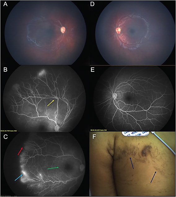
Incontinentia pigmenti (IP) is a rare X-linked dominant syndrome characterized by progressive vascular occlusive events leading to skin lesions, dental abnormalities, neurological findings, and retinal disease (Figure 5). It is thought to be caused by mutations in the IKBKG gene on the X-chromosome, thus primarily affecting females.21 Other ocular abnormalities can be seen in IP, including cataracts, strabismus, corneal opacities, uveitis, optic atrophy, and pthisis.22,23 Fluorescein angiography is essential in the staging of this disease as soon as the diagnosis is suspected or confirmed to avoid retinal detachments and blindness.24

Fluorescein Angiography Pearls
- Abnormal vascular loops and bending of vessels25
- Foveal blunting and enlarged foveal avascular zone26
- Arteriovenous shunting25
- Peripheral avascularity and leakage25
- Retinal arterial occlusions27 RP
REFERENCES
- Ghorbanian S, Jaulim A, Chatziralli IP. Diagnosis and treatment of Coats’ disease: a review of the literature. Ophthalmologica. 2012;227(4):175-182. doi:10.1159/000336906
- Villegas VM, Gold AS, Berrocal AM, Murray TG. Advanced Coats’ disease treated with intravitreal bevacizumab combined with laser vascular ablation. Clin Ophthalmol. 2014;8:973-976. Published 2014 May 16. doi:10.2147/OPTH.S62816
- Black GC, Perveen R, Bonshek R, et al. Coats’ disease of the retina (unilateral retinal telangiectasis) caused by somatic mutation in the NDP gene: a role for norrin in retinal angiogenesis. Hum Mol Genet. 1999;8(11):2031-2035. doi:10.1093/hmg/8.11.2031
- Shields JA, Shields CL, Honavar SG, Demirci H. Clinical variations and complications of Coats disease in 150 cases: the 2000 Sanford Gifford Memorial Lecture. Am J Ophthalmol. 2001;131(5):561-571. doi:10.1016/s0002-9394(00)00883-7
- Sigler EJ, Randolph JC, Calzada JI, Wilson MW, Haik BG. Current management of Coats disease. Surv Ophthalmol. 2014;59(1):30-46. doi:10.1016/j.survophthal.2013.03.007
- Hartnett ME, Penn JS. Mechanisms and management of retinopathy of prematurity. N Engl J Med. 2012;367(26):2515-2526. doi:10.1056/NEJMra1208129
- Ng EY, Lanigan B, O’Keefe M. Fundus fluorescein angiography in the screening for and management of retinopathy of prematurity. J Pediatr Ophthalmol Strabismus. 2006;43(2):85-90.
- Temkar S, Azad SV, Chawla R, et al. Ultra-widefield fundus fluorescein angiography in pediatric retinal vascular diseases. Indian J Ophthalmol. 2019;67(6):788-794. doi:10.4103/ijo.IJO_1688_18
- Early Treatment For Retinopathy Of Prematurity Cooperative Group. Revised indications for the treatment of retinopathy of prematurity: results of the early treatment for retinopathy of prematurity randomized trial. Arch Ophthalmol. 2003;121(12):1684-1694. doi:10.1001/archopht.121.12.1684
- John VJ, McClintic JI, Hess DJ, Berrocal AM. Retinopathy of prematurity versus familial exudative vitreoretinopathy: report on clinical and angiographic findings. Ophthalmic Surg Lasers Imaging Retina. 2016;47(1):14-19. doi:10.3928/23258160-20151214-02
- Mansukhani SA, Hutchinson AK, Neustein R, Schertzer J, Allen JC, Hubbard GB. Fluorescein angiography in retinopathy of prematurity: comparison of infants treated with bevacizumab to those with spontaneous regression. Ophthalmol Retina. 2019;3(5):436-443. doi:10.1016/j.oret.2019.01.016
- Hunt A, Rowe N, Lam A, Martin F. Outcomes in persistent hyperplastic primary vitreous. Br J Ophthalmol. 2005;89(7):859-863. doi:10.1136/bjo.2004.053595
- Yusuf IH, Patel CK, Salmon JF. Unilateral persistent hyperplastic primary vitreous: intensive management approach with excellent outcome beyond visual maturation. BMJ Case Rep. Published January 6, 2015. doi:10.1136/bcr-2014-206525
- Shaikh S, Trese MT. Lens-sparing vitrectomy in predominantly posterior persistent fetal vasculature syndrome in eyes with nonaxial lens opacification. Retina. 2003;23(3):330-334. doi:10.1097/00006982-200306000-00007
- Laura DM, Staropoli PC, Patel NA, et al. Widefield fluorescein angiography in the fellow eyes of patients with presumed unilateral persistent fetal vasculature. Ophthalmol Retina. Published online ahead of print July 25, 2020. doi:10.1016/j.oret.2020.07.020
- Pellegrini M, Shields CL, Arepalli S, Shields JA. Posterior tunica vasculosa lentis and “brittle star” of persistent fetal vasculature. J Pediatr Ophthalmol Strabismus. 2014;51 Online:e69-e71. Published 2014 Nov 19. doi:10.3928/01913913-20141111-01
- Pollard ZF. Persistent hyperplastic primary vitreous: diagnosis, treatment and results. Trans Am Ophthalmol Soc. 1997;95:487-549.
- Gulati N, Eagle RC Jr, Tasman W. Unoperated eyes with persistent fetal vasculature. Trans Am Ophthalmol Soc. 2003;101:59-65.
- Miyakubo H, Hashimoto K, Miyakubo S. Retinal vascular pattern in familial exudative vitreoretinopathy. Ophthalmology. 1984;91(12):1524-1530. doi:10.1016/s0161-6420(84)34119-7
- Ranchod TM, Ho LY, Drenser KA, Capone A Jr, Trese MT. Clinical presentation of familial exudative vitreoretinopathy. Ophthalmology. 2011;118(10):2070-2075. doi:10.1016/j.ophtha.2011.06.020
- Swinney CC, Han DP, Karth PA. Incontinentia pigmenti: a comprehensive review and update. Ophthalmic Surg Lasers Imaging Retina. 2015;46(6):650-657. doi:10.3928/23258160-20150610-09
- Minic S, Obradovic M, Kovacevic I, Trpinac D. Ocular anomalies in incontinentia pigmenti: literature review and meta-analysis. Srp Arh Celok Lek. 2010;138(7-8):408-413. doi:10.2298/sarh1008408m
- Goldberg MF. The blinding mechanisms of incontinentia pigmenti. Trans Am Ophthalmol Soc. 1994;92:167-179.
- Huang NT, Summers CG, McCafferty BK, Areaux RG, Koozekanani DD, Montezuma SR. Management of retinopathy in incontinentia pigmenti: a systematic review and update. J Vitreoret Dis. 2018;2:39-47.
- Liu TYA, Han IC, Goldberg MF, Linz MO, Chen CJ, Scott AW. Multimodal retinal imaging in incontinentia pigmenti including optical coherence tomography angiography: findings from an older cohort with mild phenotype. JAMA Ophthalmol. 2018;136(5):467-472. doi:10.1001/jamaophthalmol.2018.0475
- Goldberg MF. Macular vasculopathy and its evolution in incontinentia pigmenti. Trans Am Ophthalmol Soc. 1998;96:55-72.
- Cernichiaro-Espinosa LA, Patel NA, Bauer MS, et al. Revascularization after intravitreal bevacizumab and laser therapy of bilateral retinal vascular occlusions in incontinentia pigmenti (Bloch-Sulzberger syndrome). Ophthalmic Surg Lasers Imaging Retina. 2019;50(2):e33-e37. doi:10.3928/23258160-20190129-16


