


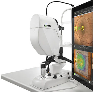
The OD-OS Inc. (Irvine, CA) Navilas Laser System 577+, a retinal photocoagulator with a digital fundus camera, has long enabled physicians to precisely deliver treatment to preplanned locations in the retina. In December, the FDA approved a compact version, the Navilas 577s, designed to extend the benefits of its Laser System 577+ to practices and clinics of all sizes.
The Navilas system is indicated for all retinal diseases treated with laser. Studies have shown that the Navilas can be a significant partner in reducing the number of anti-VEGF injections.
David M. Brown, MD, clinical professor of ophthalmology at Baylor College of Medicine in Houston, TX, has been using the Navilas technology both in day-to-day clinic operations and as part of a randomized, clinical trial for three years. The system “eliminates human variability for both patients and surgeons,” he says. “Analogous to eye-tracked devices used in refractive surgery, the Navilas laser system delivers the prescribed treatment to patients regardless of their eye movements.”
The technology, he continues, enables physicians to treat pathology that is only visible on fluorescein angiography (FA), indocyanine green angiography, or optical coherence tomography (OCT). And it does so, he adds, “in a precise, controlled fashion.”
Dr. Brown notes that more than 50% of diabetic and retinal vein occlusion macroaneurysms (MAs) can only be seen on FA; besides these, the Navilas allows treatment on visible MAs. “Collateral damage that is inevitable with manual lasers doesn’t occur, allowing surgeons to aggressively target pathologies near the fovea that would be too dangerous to work on otherwise,” he says.
HOW IT WORKS
Navilas uses the OD-OS proprietary reflex-free fundus imaging system to acquire high-resolution video and still images of the patient’s retina. This can be done using true color or infrared light. The system allows physicians to preplan the treatment based on the system’s color fundus images or on additional images provided by their own OCT and FA instruments. The system automatically registers these images to the live fundus.
The physician can then quickly create a treatment plan by placing individual spots or grids of any desired shape on these diagnostic images. During the treatment phase, the computer guides the laser to all preplanned locations, recording each successful laser application. This process lets the physician focus on delivering high-quality treatment, while the system stabilizes the laser on the patient’s retina, compensating for eye movements. As an added benefit, the combination of advanced eye tracking and infrared illumination allows for focal treatments without the use of contact lenses.
CLINICAL APPLICATIONS
The Navilas system is indicated for use in photocoagulation for the treatment of clinically significant diabetic macular edema, proliferative diabetic retinopathy (panretinal photocoagulation), subretinal (choroidal) neovascularization, central and branch retinal venous occlusion, lattice degeneration, retinal tears, and detachments. It is also indicated for retinal imaging, including color and infrared imaging, and for aiding in the diagnosis and treatment of ocular pathology in the posterior segment of the eye.
IMPROVING PATIENT OUTCOMES
The need for anesthesia is reduced because no contact lens needs to be worn during focal treatments.
Both focal and panretinal photocoagulation treatments can be completed using infrared illumination, which results in a significant reduction in pain perceived by the patient, according to the company website. Also, the increased speed of Navilas results in shorter treatments.
In the short term, patients benefit from 30% increased accuracy compared to conventional lasers.1 Furthermore, many treatments can be completed in a single session, attaining a faster and further reduction in central retinal thickness and 40% fewer retreatments compared to conventional laser delivery.2
If used in conjunction with anti-VEGF injections, Navilas significantly lowers the treatment burden, reducing the amount of injections required by 75% over traditional monotherapy, supporting better long-term outcomes.3 The Navilas-guided laser has also led to fewer pharmacotherapy injections, as suggested by several studies, Dr. Brown adds. The TREX DME study,4 underway at multiple centers, is expected to provide guidance on the magnitude and duration of this response.
USER-FRIENDLY PLANNING AND TREATMENT
The software in the newly approved Navilas 577s makes the planning and treatment phase seamless, with quicker treatment planning than with the original device, Dr. Brown says. “The smaller optical head [compared to the Navilas 577+] allows surgeons to see the patient better, ensuring improved communication.” The 577s’ technology enables high-resolution imaging of the retina with a large field of view throughout treatment, resulting in enhanced visualization compared to conventional slit-lamp-based lasers, which only partially visualize the retina. Physicians can access functions directly on the touchscreen, as well as by using buttons on the base and joystick. During the learning phase, both the trainer and trainee can watch the screen, instead of viewing through an observation tube. This enables direct communication and gives the trainer the opportunity to identify potential mistakes before laser energy is applied.
The navigation also allows physicians to block sensitive areas with exclusion zones and to digitally document every laser spot. At any point during or after treatment, colleagues and even patients can be involved in the treatment by reviewing the plan and spot locations. The final treatment result can be easily printed or shared electronically with referring physicians. RP
REFERENCES
- Kozak I, Oster SF, Cortes MA, et al. Clinical evaluation and treatment accuracy in diabetic macular edema using navigated laser photocoagulator NAVILAS. Ophthalmology. 2011;118(6):1119-1124.
- Neubauer AS, Langer J, Liegl R, et al. Navigated macular laser decreases retreatment rate for diabetic macular edema: a comparison with conventional macular laser. Clin Ophthalmol. 2013;7:121-128.
- Liegl R, Langer J, Seidensticker F, et al. Comparative evaluation of combined navigated laser photocoagulation and intravitreal ranibizumab in the treatment of diabetic macular edema. PLoS One. 2014;9(12):e113981.
- Payne JF, Wykoff CC, Clark WL, Bruce BB, Boyer DS, Brown DM; TREX-DME Study Group. Randomized trial of treat and extend Ranibizumab with and without navigated laser for diabetic macular edema: TREX-DME 1 year outcomes. Ophthalmology. 2017;124(1):74-81.