PHOTO ESSAY
Blind Spots/White Dots
NANCY KUNJUKUNJU, MD
The patient is a 30-year-old male, who had a 2-day history of "blind spots" in his left eye. His past ocular history is significant for myopia and hard contact lens use. Past medical history and family history were not significant. He took no medications and reported no allergies. The patient had an extensive travel history; a thorough review of systems was otherwise negative. Extensive lab work was negative for anything unusual.
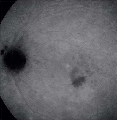
On initial exam, his vision was 20/25 in both eyes, anterior segment was benign and there was no evidence of vitreal cell. Inferiotemporal to the fovea of his left eye, he had a hypopigmented lesion deep to the retinal vessels (Figures 1A and 1B). On fluorescein angiography (FA), the hypofluorescent lesion appeared to be a cluster of hyperfluorescent dots (Figure 2), which were more apparent than on clinical examination. These dots appeared to stain slightly during late-phase angiography (Figure 3). Early-phase indocyanine green angiography (ICG) was unremarkable; however, during late-phase angiography, there were small hypofluorescent dots overlying larger hypofluorescent spots (Figure 4). There was also a lacy, irregular ring of hypofluorescence at the margin of the optic disc. The hypofluorescent spots appeared more numerous than was apparent on either fluorescein or on clinical exam. Microperimetry was performed. The imaged blind spots on microperimetry (Figure 5) corresponded to the hypopigmented areas noted on ICG. One week after the initial visit, multiple hyperfluorescent white dots appeared on the FA (Figure 6); during this visit, the patient had vitritis that resolved spontaneously within a few days. Repeat microperimetry (Figure 7A) and ICG (Figure 7B) showed improvement 6 weeks after the initial visit and after a course of steroids. Imaging supports a diagnosis of multiple evanescent white dot syndrome (MEWDS). RP
| Nancy Kunjukunju ia a retinal fellow at the Retina and Vitreous Center in Ashland, OR. She reports no financial interest in any products mentioned here. She can be reached via e-mail at nkunjukunju@gmail.com. |

Figure 1A. Fundus photo showing clinically apparent hypopigmented lesion (initial visit).

Figure 1B. Red-free photo of the lesion (initial visit).

Figure 2. Laminar phase angiography showing the lesion as a cluster of white dots (arrow).

Figure 3. Late-phase angiography showing staining of the white dots (arrow).

Figure 4. Late-phase ICG showing multiple areas of hypofluorescence, particularly surrounding the optic nerve.

Figure 5. "Blind spots" on microperimetry that parallel the hypopigmented areas noted on ICG.

Figure 6. One week later, there are multiple white dots (arrows).

|

|
Figure 7A and B. Microperimetry and ICG taken 6 weeks after the initial visit and a course of steroids. The "blind spots" appeared to be resolving on microperimetry, which corresponded to an improved ICG.


