|
|
|
Figure 1. Fundus photograph (upper left) and fluorescein angiogram before the subthreshold micropulse diode laser treatment. Fluorescein angiogram shows early and late leaking aneurysms. |
feature
Diode Subthreshold Micropulse Laser for Diabetic Macular Edema
A
new laser modality has been introduced into clinical practice.
NEELAKSHI BHAGAT, MD, MPH • MARCO
A. ZARBIN, MD, PhD
The current treatment for clinically significant diabetic macular edema (CSME) is focal or grid laser photocoagulation as outlined by the Early Treatment Diabetic Retinopathy Study (ETDRS).1 Although this treatment improves the visual prognosis of patients with CSME, it is not a sight-restoring therapy. Only 3% of patients have any moderate improvement in vision after conventional laser treatment for CSME. A new laser modality, the micropulse laser, has been introduced recently into clinical practice.2-4 Subthreshold micropulse diode laser photocoagulation (SMDLP) is designed to produce multiple short exposure burns that can be focused on a very small area of targeted tissue. Physical characteristics of the light absorbance of the retina and retinal pigment epithelium (RPE) are such that these extremely short pulses of laser energy target thermal changes to the RPE with minimal diffusion of energy to the overlying neurosensory retina, thus minimizing unnecessary retinal damage.5 In contrast, conventional laser treatment damages both the RPE and overlying retina. These properties of micropulse laser have led to exploration of its use in the treatment of macular edema.
CASE REPORT
|
|
|
Figure 2. OCT before the subthreshold micropulse diode laser treatment showing mild retinal thickening in the perifoveal and foveal areas. |
A 59-year-old man with noninsulin diabetes mellitus, presented
for evaluation of diabetic retinopathy. His best-corrected visual acuity (VA) was
20/20 in both eyes. Slit-lamp biomicroscopy of the anterior segment was normal.
Fundus exam disclosed diabetic macular edema (DME) in the left eye, which was not
clinically significant. At follow-up 3 months later VA remained 20/20 in the left
eye, but fundus exam revealed progression of the edema within
1-disc diameter
of the foveal avascular zone (FAZ) center and the presence of CSME. Retinal thickening
was present mostly at the temporal and inferonasal edge of the FAZ, ~1.5 mm from
the macular center. Focal laser photocoagulation for CSME was recommended.6
The fluorescein angiogram demonstrated a normal FAZ with leaking microaneurysms in the parafoveal area (Figure 1). Optical coherence tomography (OCT) (Stratus OCT,
Carl Zeiss Meditec, Dublin, Calif) revealed minimal thickening within the 3.45-mm diameter area centered at the foveola (Figure 2). Subthreshold micropulse diode laser photocoagulation was recommended due to the proximity of the retinal edema to the fovea in a patient with an ETDRS VA of 20/20.
|
|
|
Figure 3. Color photograph and fluorescein angiogram 1 day after the second subthreshold micropulse diode laser (SMDLP) treatment. Most of the 1 day old SMDLP spots (arrows in the 6 min 35.2 sec frame) are seen as hyperfluorescent areas in the mid frame (2 min 41.0 sec) that stain markedly in the late frame (6 min 35.2 sec). The areas of blocked fluorescence (arrows) seen in the early frame (44.8 sec) are the old SMDLP marks from the first treatment performed 3 months before. |
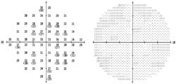
Focal laser was performed using diode micropulse laser in a subthreshold manner within 3 mm of the fovea mostly temporal and inferonasal to the fovea not specifically targeting the microaneurysms. The following settings were used: cycle 15%; power 750 milliwatts (mW); envelope duration 0.150 s, 75 μm spot size, #32 spots. The spots were invisible clinically. The patient returned 4 months later with a stable ETDRS VA of 20/20. The macular edema had decreased, but residual edema was present. The patient underwent another round of diode subthreshold micropulse laser therapy. The settings were: cycle 15%; power 750 mW; envelope duration 0.15 s; spot size 75 μm and #22 spots in the parafoveal area within a 2-disc diameter area centered at the fovea. The laser spots were again invisible. A follow-up fluorescein angiogram the next day revealed hyperfluorescence of many (but not all) laser spots delivered the day before (Figure 3). The patient returned 4 months later (8 months after the first laser treatment). The macular edema had resolved. Optical coherence tomography revealed much decreased retinal thickness within the 3 mm radius of the FAZ center (Figure 4). Fundus exam disclosed no laser spots with biomicroscopy, but the fluorescein angiogram did reveal some RPE changes at the site of the laser spots (Figure 5). Humphrey visual field testing and scanning laser ophthalmoscope (SLO) microperimetry disclosed no significant changes in paracentral scotomata (Figures 6 and 7).
DISCUSSION
Mechanism of Laser Action
The exact mechanism of action of laser is unknown. The RPE proliferates and migrates after laser photocoagulation, and the outer blood-retinal barrier may undergo both anatomic and functional restoration.5-8 In animal and human RPE cell culture experiments, laser photocoagulation promotes production of TGF-b by RPE cells that antagonizes the vascular permeability-inducing effects of vascular endothelial growth factor.9 Experimental findings have shown that photocoagulation and stimulation of the RPE layer alone, without causing a destruction of the overlying retinal structure, is enough to mediate a beneficial effect in the retina. Retinal pigment epithelium defects after selective subthreshold laser burns are covered by a new population of RPE cells.5 With the availability of the micropulse mode of laser and with subthreshold intensity, we may be able to limit the thermal damage solely to the RPE layer.
Micropulse Diode Laser Photocoagulation
|
|
|
Figure 4. OCT after 2 subthreshold micropulse diode laser treatments (8 months after the first treatment). The central foveal thickness has decreased from 230 μm to 207 μm. The perifoveal thickness within the 3.45 mm radius (within the 2 rings around the fovea) has also decreased when compared to the prelaser OCT (Figure 2). |
The micropulse laser is delivered in very short "bursts" or pulses. Heat generated with each short pulse is small, such that it minimally diffuses to the adjoining tissue. The target tissue, RPE, is affected while the choroid and neurosensory retina is spared, leading to fewer complications, in principle. The laser spots are also delivered in a subthreshold manner, which means that the laser treatment spots are invisible on clinical examination. Thus, much less energy is delivered with each burst of subthreshold micropulse laser compared to conventional laser photocoagulation.
Complications due to thermal damage to the retina and/or choroid (eg, symptomatic paracentral scotomata, choroidal neovascularization) are minimized with laser pulses of very short duration that affect the RPE alone with little effect on the photoreceptors or choriocapillaris.10,11 Complications such as foveal distortion, accidental foveal burns, subretinal fibrosis, decreased contrast sensitivity, blood vessel perforation, and chorioretinal anastomosis correlate directly with the energy delivered per laser spot. Use of subthreshold micropulse laser should minimize these undesired effects. In our patient, no new relative or absolute scotomata were noted on Humphrey visual field central -10 (Figure 6) or with SLO microperimetry (Figure 7).
The technique of SMDLP has been described previously.3 We use the diode 810 nm micropulse laser with the following settings: an "on" time of 300 μs, an "off" time of 1700 μs (15% cycle), and an envelope duration of 0.3 s–0.4 s. A test burn is delivered outside the arcades using a power intensity such that a faint white burn is seen. Once the parameters of the test burn are defined, the power and the envelope duration are reduced by 50%. Typically, the parameters of the subthreshold spot (after the test dose) are: 15% cycle, power 750 mW, and envelope duration 0.15 s.
|
|
|
Figure 5. Fundus photograph and fluorescein angiogram after 2 subthreshold micropulse diode laser treatments (8 months after the first treatment). |
This case demonstrates the use of SMDLP in the treatment of DME. It is very useful in patients with edema very close to and within the foveal avascular zone. In our patient, the CSME resolved, and the VA has been stable at 20/20.
REFERENCES
1. ETDRS Research group. Photocoagulation for diabetic maculae edema: ETDRS Report No. 1. Arch of Ophthalmol. 1985;103:1796-1806.
2. Bhagat N, Zarbin MA. Diode Subthreshold Laser for DME. Retinal Insider. 2002;83-86.
3. Bhagat N, Zarbin MA. Use of diode subthreshold micropulse laser for treating diabetic macular edema. Contemporary Ophthalmology. 2004;3: 1-5.
4. Laursen ML, Moeller F, Saunders B, Sjoelie AK. Subthreshold micropulse diode laser treatment in diabetic macular edema. Br J Ophthalmol. 2004;88:1173-1179.
5. Roider J, Michaud N, Flotte TEA. Response of the RPE to selective photocoagulation of the RPE by repetitive short laser pulses. Arch Ophthalmol. 1992;110:1786-1792.
6. Ferris FL 3rd. Davis MD. Treating 20/20 eyes with diabetic macular edema. Archives of Ophthalmol.1999;117:675-676.
|
|
|
Figure 6. Humphrey VF central -10 (pre- and
8 months-post laser treatment). No worsening of relative or absolute scotomata is
noted. Top – prelaser with CSME; Bottom – after both subthreshold micropulse
diode laser treatments, resolved CSME. |
7. Lee CM, Olk J. Modified grid laser photocoagulation for diffuse diabetic macular edema. Ophthalmology. 1991;98:1594-1602.
8. Bresnick GH. Diabetic maculopathy; a critical review highlighting diffuse macular edema. Ophthalmology. 1983;90:1301-1317.
9. Tolentino MJ, Mcleod DS, Taomoto M, Otsuji T, Adamis AP, Lutty GA. Pathologic features of vascular endothelial growth factor-induced retinopathy in the nonhuman primate. Am J Ophthalmol. 2002;133:373-385.
10. Toth CA, Narayan DG, Cain CP, DiCarlo CD, Rockwell B, Roach WP. Histopathology of ultrashort pulse laser retinal lesions. Invest Ophthalmol Vis Sci. 1996;37:S694.
11. Chong LP, Soriano D, Ramos AR. Sublethal laser damage to the retinal pigment epithelium by micro-pulse diode laser in primate eye. Invest Ophthalmol Vis Sci. 1996;37:S694.
Neelakshi Bhagat, MD, MPH, is assistant professor of Ophthalmology and director, Vitreo-retinal and Macular Surgery at the Institute of Ophthalmology and Visual Science, New Jersey Medical School. Marco A. Zarbin, MD, PhD, FACS, is professor and chair of the Institute of Ophthalmology and Visual Science, New Jersey Medical School. Neither author has any financial interest in the information presented in this article. Dr. Bhagat can be reached by e-mail at bhagatne@umdnj.edu and Dr. Zarbin can be reached by e-mail at zarbin@earthlink.net. This work was supported by: Research to Prevent Blindness Inc., the Lions Eye Research Foundation of New Jersey, and the Eye Institute of New Jersey.
|
|
|
Figure 7. SLO microperimetry. Top: prelaser; Bottom: No absolute or relative scotomata are noted 8 months after 2 subthreshold micropulse diode laser treatments. |


