feature
State-of-the-Art
Imaging Technology
A
review of new advances in diagnostic retinal imaging.
COMPILED BY THE RETINAL
PHYSICIAN EDITORIAL STAFF
Recent advances in the field of retinal imaging have furthered ophthalmologists' understanding of retinal anatomy. With today's various technologies, a comprehensive view of the posterior segment can be realized. This capability is invaluable in diagnosing and monitoring retinal disorders.
Here are several of the contemporary retinal diagnostic imaging products on the market.
|
|
|
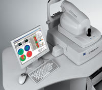
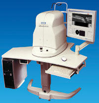
The Stratus has been used in vital clinical studies. |
►Stratus OCT (Carl Zeiss Meditec, Dublin, Calif). The Stratus OCT provides quantitative and qualitative assessment of the retina. With the Stratus OCT Review Software, physicians can now import, view, analyze, and manage the Stratus OCT exam data on a personal computer.
Dana Deupree, MD, Palm Harbor, Fla, uses the OCT for determining whether a patient needs a procedure as well as planning surgical approaches. For example, he uses the Stratus in diabetics to find tight hyaloid membranes to gauge if they are creating macular edema, which might be an indication for surgery. "You can see the actual anatomy and how it is oriented to various membranes, and how they are affecting the central macula," he notes. Dr. Deupree also uses the OCT as a barometer to judge whether a treatment is efficacious. "It gives you an objective measurement as to the effectiveness of the current treatment regimen; seeing the actual histology and being able to apply an objective measurement to that as a baseline or benchmark allows me to monitor changes," explains Dr. Deupree.
|
|
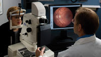
| Escalon's product has single or dual cameras. |
►High-Resolution Imaging System (Escalon Digital Solutions, Wayne, Pa). This system has 6 Mega-Pixel, contains full-frame sensors, maximum sensitivity,
optimalcolor imaging, and fluorescein angiography (FA). Image processing algorithms specifically designed for fundus imaging optimize image quality and provide instantaneous image capture. System software includes patient database, calibrated measurement functions, an auto montage imaging feature, and networking and connectivity options.
Thomas N. Fleming, MD, Belleville, Ill, uses the system primarily for age-related macular degeneration (AMD) and diabetic retinopathy (DR) patients. He says the system is good at picking up capillaries in and around the fovea for DR. "I like the resolution of the pictures," says Dr. Fleming. In addition to those indications, he uses it for branch retinal vein occlusion patients and to screen for cystoid macular edema in post-cataract eyes.
Dennis Zukosky, ophthalmic photographer in Dr. Fleming's practice, says, "It's a complete system; it gives the doctor what he needs and it is flexible."
|
|
|
The HRA 2 is a confocal laser imaging system. |
►HRA2 (Heidelberg, Vista Calif). The Heidelberg Retina Angiograph 2 system can change a series of optical sections into a layered, 3-D image of the retina and enables standard high-resolution, high-contract FA and indocyanine green angiography (ICGA). It also performs autofluorescense (488 nm) imaging using the normal FA settings, but without the injection of fluorescein. Scott Cousins, MD, Durham, NC, uses the HRA2 as a diagnostic adjunct for identifying choroidal neovascularization (CNV) in pigment epithelial detachments; distinguishing between CNV and retinal angiomatous proliferation (RAP); and finding what he deems are "mature vascular complexes" in CNV. This term refers to a mature new vessel complex that has been in the posterior segment for sometime and has a well-defined feeder vessel and arterioles perfusing the lesion.
Dr.
Cousins finds the scanning laser system useful as it avoids the "artifacts of scattering,"
which allows a much better signal. Another important feature he utilizes is the
HRA2's dynamic, high-speed movies, which can capture up to 16 frames per second
during FAs and/or ICGAs.
Dr. Cousins says the beginning of imaging scans is
paramount and is where the most useful diagnostic information is located —
like blood-flow characteristics, for example.
"You can only get that information if you have a movie or a video," explains Dr. Cousins. "Whereas with a standard ICG, you are capturing 1 frame every 2 or 3 seconds, and physicians have a tendency to focus on the late frames."
|
|
|
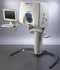
The optomap FA images over 80% of the retina. |
►Optomap FA Digital Ultra-widefield Angiography (Optos, Marlborough, Mass). This product produces real-time, high-resolution angiographic images, and allows simultaneous evaluation of the peripheral and central retina. It offers information to help monitor and diagnose eye conditions and to assist in treatment determinations. Visualization of the periphery may help early identification of patients at higher risk of disease progression.
Steven Schwartz, MD, Los Angeles, has had the benefit of experience with the optomap FA as his practice has been working with the prototype. "We have found it useful for all retinal vascular disease," says Dr. Schwartz. He also credits its ease of use.
Within his practice, he and fellow surgeons have preferred using it to conventional angiography. Dr. Schwartz also credits the widefield view, and says it allows physicians to get a more comprehensive view of the retina. He points specifically to its ability to detect retinal non-anterior perfusion early.
While he points out this is a new technology, he is optimistic about the optomap FA in the long view. "It has the potential to have a big impact [in diagnosing retinal disease,"] says Dr. Schwartz.
|
|
|
The OCT/SLO has a single super-luminescent diode. |
►OCT/SLO (OTI, Toronto, Ontario). OCT and SLO images of the fundus are produced simultaneously through the same optics. The OCT and SLO images correspond pixel to pixel. OCT images can be acquired either as B-Scan of the retina and/or as a parallel sectional stack of C-Scan images at variable depths.
Richard Rosen, MD, New York, utilizes the OCT/SLO primarily for AMD and DR screenings. He finds it to be effective with its ability to combine B- and C-scans, and it allows him to obtain a comprehensive view of the posterior segment from front to back."You can correlate the surface of the eye, with what is going on inside," says Dr. Rosen.
Dr. Rosen also likes how the OCT/SLO images are scanned compared to other diagnostic products. "When you scan parallel to the surface, you have better continuity." This allows him to locate more "subtle pathologies."
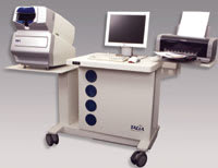
►Retinal Thickness Analysis (RTA 5) (Talia/Marco, Jacksonville, Fla). Outputs include thickness and topography analysis, interactive 3-D imaging, retinal nerve fiber layer measurement, optical cross sections, digital fundus imaging, and an overlay of a 2-D thickness map onto a patient's digital fundus image. The RTA 5 offers its "Vision Wellness" report for baseline exams. A new 'Dynamic 3-D Anatomy Imager' program allows continuous visualization and quantitative analysis through each cross-sectional slit image, across any scanned field.
|
|
| The RTA 5 is made by Talia and sold by Marco. |
Sara Krupsky, MD, Rehovot, Israel, uses the RTA 5 to detect and monitor a variety of ailments. "The RTA 5's high-quality fundus photography with thickness mapping makes it an excellent tool to detect DR and follow these patients after treatment." She also uses it identify conversion of dry to wet AMD and to monitor CNV resolution following treatment. As Dr. Krupsky has the RTA in her practice, she uses it as an indicator for pathological changes (subretinal fluid, pigment epithelial detachment, macular edema) thus avoiding repeated and invasive FA testing.
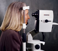
►TRC-NW6S (Topcon, Paramus, NJ). This non-mydriatic fundus camera's 9 internal fixation LEDs help document a wide-aspect view of retina. Angle data and OS/OD information transfers automatically to IMAGEnet software. IMAGEnet provides a myriad of software features to make the images useful in managing patient compliance and monitoring disease progression.
Douglas Kaplan, MD, Chicago, uses the TRC-NW6S to monitor progression of dry AMD, document and treat DR, and establish baseline imaging in retinal/choroidal lesions. Dr. Kaplan also shows his AMD and DR patients their images from the TRC-NW6S. He believes it is a beneficial tool in helping these patients understand their diseases and participate in their own care.
|
|
|
Topcon's TRC-NW6S offers 45Þand 30Þ fields. |
"When they can see what is happening to their eyes, it really boosts their knowledge and compliance," he says.
CONTINUOUS INNOVATION
Today's systems provide a more comprehensive view of the posterior segment. As the technology continuously improves, it will further aid physicians with diagnostic challenges in helping to identify hidden pathologies.


