Wet AMD Drug
Delivery Options
To reach treatment safety, efficacy, and acceptance, the best delivery methods must be sought.
MICHAEL J. COONEY, MD
Ophthalmic drug delivery is back in the spotlight due to the excitement surrounding new and emerging treatments for posterior segment disease. Currently, these treatments are delivered in a variety of ways, including systemically, by intravitreal injection, via sustained-release implants, and posterior juxtascleral depot (PJD). As more treatments become available, patient acceptance and safety issues will play a role in which treatments are widely used.
Delivery methods for age-related macular degeneration (AMD) treatments are now gaining attention for good reason. An estimated 15 million people have AMD in the United States, and these numbers will rise sharply with the dramatic demographic right-shift expected in the next 20 years.1 While only 10% of AMD cases are of the wet choroidal neovascularization (CNV) variation, this type accounts for 90% of the vision loss in AMD. Thus, wet AMD is an urgent medical need that has several pharmaceutical companies working on new treatments.
This article will review drug-delivery methods that are currently in use or under investigation for use in wet AMD.
INTRAVITREAL INJECTION
Until rather recently, intravitreal injection was a relatively obscure drug delivery method that was used for treating endopthalmitis, retinal detachment, and cytomegalovirus retinitis. However, in the past several years, intravitreal injection has seen new life in treating various retinal diseases.
Triamcinolone acetonide (Kenalog) injection, which is increasingly given as adjunctive therapy with verteporfin (Visudyne) photodynamic therapy, has shown encouraging clinical results in small uncontrolled studies. Clinical trials are ongoing to provide conclusive data regarding adjunctive intravitreal triamcinolone in photodynamic therapy.
Pegaptanib sodium (Macugen), like triamcinolone, is also delivered via intravitreal injection. It received FDA approval in December 2004, and became commercially available in January of this year. Pegaptanib sodium is an aptamer that selectively binds VEGF, one of the many factors that can trigger angiogenesis in the posterior segment. Pegaptanib sodium is injected every 6 weeks and positive efficacy has been reported across all CNV lesion subtypes.2-4
Delivery of a therapeutic agent with an intravitreal injection is direct and reliable, and controlled clinical studies for drugs have improved the safety profile of the procedure.
However, intravitreal injections are associated with some complications. The 54-week phase 2/3 studies for pegaptanib sodium reported endophthalmitis, traumatic cataract, and retinal detachment as the most common potential risks (Table).4
|
Table. Most Common Complications of Pegaptanib Sodium From Macugen Pivotal Clinical Trials (54 weeks) |
||
| Adverse Event | Number #009; of Patients | Patient Percentages #009;per injection |
| Endopthalmitis | 12 | 1.3 (0.16) |
| Traumatic Cataract | 5 | 0.6 (0.07) |
| Retinal Detachment | 5 | 0.6 (0.07) |
| Source: FDA Macugen Advisory Board Meeting; Rockville, MD, August 27, 2004 | ||
Twelve cases of endopthalmitis were reported in the 54-week data from the pivotal studies; however, including all studies, 16 cases were reported at 54 weeks. As of the FDA Advisory Committee meeting, a total of 18 patients had developed endopthalmitis: 16 cases were reported before a protocol amendment was instituted and 2 cases were reported after the amendment.
Prior to the protocol amendment, the injection procedure was performed after application of 23 drops of 10% povidone iodine or 1 drop of topical antibiotic. Subsequent to the protocol amendment, patients were asked to administer topical antibiotic drops for 3 days prior to surgery or were flushed with povidone iodine prior to injection. The protocol amendment also required the use of a sterile surgical drape and lid speculum.
|
|
|
|
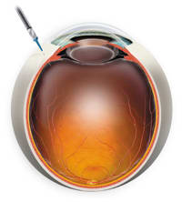
Figure 1. A 27 g needle 3.5 4.0 mm posterior to the limbus. |
|
Intravitreal injections are generally performed with a lid speculum in place. A 27 g needle is typically used for the procedure with an injection site 3.5 4.0 mm posterior to the limbus (Figure 1).
TOPICAL THERAPY
Although simple, topical delivery is of limited value in treating posterior-segment disease. As much as 90% of a topically applied drug is immediately flushed away by the blink mechanism and lacrimal system during delivery. The successful absorption of the drug through the anterior segment tissues requires a complex balance of hydrophilic and hydrophobic drug properties. Furthermore, once absorbed into the front of the eye, only a small percentage of drug becomes bioavailable to the posterior segment.5
Despite these challenges, Othera Pharmaceuticals is developing a topical drug for AMD treatment and arresting cataract progression. The topical solution OT551 is a new chemical entity derived from tempol-H that has shown bioavailability in the posterior chamber and retina in animal models.
OT551 is a free radical scavenger that might prevent retinal damage due to oxidative stress, which is thought to be a part of the early stages of AMD. No clinical data is available on this compound at this time and we await the results of clinical studies.
POSTERIOR JUXTASCLERAL DEPOT
This method is a novel technique that was developed by Alcon for the administration of anecortave acetate (Retaane). The New Drug Application for anecortave acetate was completed in December 2004 and Alcon anticipates FDA approval by mid year 2005.
Posterior juxtascleral depot requires inserting a blunt, curved cannula into a conjunctival incision (1.0 1.5 mm) located 8 mm posterior to the superotemporal limbus.
The curved cannula follows the surface of the sclera, without puncturing the globe, and anecortave acetate is injected in the juxtascleral space overlying the macula. Anecortave acetate binds to the sclera and delivers drug to the choroid over a 6-month period. No serious treatment-related effects have been reported to date due to the PJD technique.
As reported in the clinical trial results for anecortave acetate at the 2004 American Academy of Ophthalmology (AAO) Meeting in New Orleans, drug reflux may affect its efficacy. Reflux occurs when the drug flows back out of the incision made at the insertion site.
|
|
|
|
Figure 2. A CPD and cannula in place for PJD procedure. |
|
To address this problem, Alcon developed a counter pressure device. This simple device, when used during the PJD procedure, stops reflux and does not add significant complications to the overall procedure (Figure 2). Alcon initiated a pharmacokinetic study which tested the efficacy of the device and recent results confirmed its value.
INTRAVENOUS
Verteporfin has been approved since 2000 for subfoveal predominantly classic CNV secondary to AMD. This drug can be classified as a systemic drug because Verteporfin is administered intravenously and complexes with low density lipoproteins which are prevalent in new vasculature.6
Another intravenously delivered, light-activated drug in development is tin ethyl etiopurpurin (Photrex formerly known as SnET2). This drug functions largely by the same mechanism as verteporfin. The FDA issued an approvable letter for tin ethyl etiopurpurin in September 2004, requesting additional clinical investigation.
An intravenous drug under development for CNV due to myopic macular degeneration is Combretastatin A4 phosphate (CA4P). It is concurrently under investigation for the treatment of certain types of cancer. Combretastatin A4 phosphate is an antivascular tubulin-binding agent, which is derived from a South African willow tree.
The drug suppresses neovascularization while permitting the development of normal retinal vasculature and it has shown promising results in preclinical and clinical models.7 Combretastatin A4 phosphate was approved by the FDA for investigational use in CNV secondary to myopic macular degeneration in November 2004.
IMPLANTS
Effective drug delivery to the posterior segment can also be achieved using intravitreal implants. These implants release a therapeutic level of drug over a specified period of time, avoiding the need for frequent retreatments. Fluocinolone (Retisert) and dexamethasone (Posurdex) are 2 examples of implants that are undergoing clinical investigation for posterior-segment diseases.
Bausch & Lomb filed for approval of fluocinolone in December 2004 for noninfectious uveitis, and approval of the device is anticipated in 2005. The device is implanted in the operating room and releases the drug for up to 3 years. Clinical studies demonstrate that the uveitis recurrence rate is significantly reduced in the fluocinolone implant-treated eyes compared with control eyes. Additional clinical results for fluocinolone will be released in late 2005.
Posurdex is a biodegradable polymer implant that releases dexamethasone as the polymer degrades. Posurdex is under clinical investigation for the treatment of persistent macular edema secondary to diabetic retinopathy, uveitis, and vein occlusion.
VIRUSES AND GENETIC TECHNOLOGY
Pigment epithelium derived factor (PEDF) is a naturally occurring antiangiogenic molecule. Preliminary evidence shows that PEDF inhibits fibroblast growth factor, platelet-derived growth factor, interleukin-8, and vascular endothelial growth factor.
Technology developed by GenVec, Inc. packages the gene for PEDF into a viral vector (AdPEDF), which is delivered via an intravitreal injection. After delivery, the PEDF gene is incorporated into cells within the posterior segment and PEDF proteins are synthesized.
In a small phase 1 study, in which 27 subjects received injections of AdPEDF, the treatment was well tolerated up to 1x105.9 particle units. High levels of PEDF were found in the eye 24 hours after injection and decreased with time. PEDF levels were still measurable after 14 days.8
CONCLUSION
Development of new pharmacological treatments for CNV and other ocular diseases has refocused attention on drug-delivery methods. Risks and benefits of different delivery methods will likely carry increased weight in patient and clinician treatment decisions as more treatment options become available. Progress has been made in finding the ideal combination of clinical efficacy, duration of action and minimal complications, but more research and innovation will inevitably lead to better patient care.
Address correspondence to: Michael J. Cooney, MD, Assistant Professor of Ophthalmology and Director of the Medical Retina Service, Duke University Eye Center, Box 3802, DUMC, Durham, NC 27710, Telephone: (919) 684-3090, Fax: (919) 681-6474, E-mail: coone004@mc.duke.edu.
From Retina Service, Duke University Eye Center, Durham, NC. Dr. Cooney is a
consultant for Bausch & Lomb.
REFERENCES
1. Klein R, Klein BE, Linton KL. Prevalence of age-related maculopathy. The Beaver Dam Eye Study. Ophthalmology. 1992;99:933-943.
2. The Eyetech Study Group. Preclinical and phase 1a clinical evaluation of an anti-VEGF pegylated aptamer (EYE001) for the treatment of exudative age-related macular degeneration. Retina. 2002;22:143-152.
3. Eyetech Study Group. Anti-vascular endothelial growth factor therapy for subfoveal choroidal neovascularization secondary to age-related macular degeneration: phase II study results. Ophthalmology. 2003;110(5): 979-986.
4. FDA Macugen Advisory Board Meeting; Rockville, MD, August 27, 2004.
5. Worakul, Nimit et al. Ocular Pharmacokinetics. In: Albert D, ed. Principles and Practice of Ophthalmology, 2nd ed. Philadelphia, PA: W.B. Saunders Company; 2000;202-211.
6. TAP Study Group. Photodynamic therapy of subfoveal choroidal neovascularization in age-related macular degeneration with verteporfin: one-year results of 2 randomized clinical trials. Archives of Ophthal. 1999;117(10):1329.
7. Nambu H, NambuR, Melia M, Campochiaro PA. Combretastatin A-4 phosphate suppresses development and induces regression of choroidal neovascularization. Invest Ophthalmol Vis Sci. 2003;44(8):3650-3655.
8. Holtz ER. American Academy of Ophthalmology Retina Subspecialty Day; New Orleans, LA, October 22, 2004.


