Imaging
Assessing RPE Function with Autofluorescence
|

|
|
|
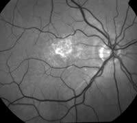
The digital color fundus image and the red-free image show fibrin and atrophic changes in this patient's macular area. |
|

|
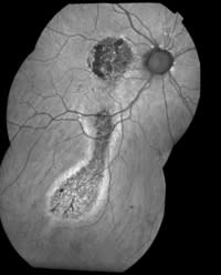
60-year-old female with chronic central serous chorioretinopathy
Autofluorescence imaging of the ocular fundus relies on the stimulated emission of light from molecules, chiefly lipofuscin, in the retinal pigment epithelium (RPE). Lipofuscin is a diverse group of molecular species, yellow to brown in color, that accumulates in all post-mitotic cells from the oxidative breakdown and rearrangement of a number of different molecules, including polyunsaturated fatty acids and proteins.
The RPE has the important job of phagocytizing photoreceptor outer segments, which contain retinols. The main component of lipofuscin in RPE cells is a molecule called A2E, which is formed from two molecules of transretinal and one molecule of phosphatidlyethanolamine. By age 70 an average RPE cell will have phagocytized 3 billion outer segments, and more than 25% of the cell volume will be occupied with lipofuscin.
One intriguing aspect of lipofuscin in general is that it can be made to fluoresce. In ophthalmology we call this type of fluorescence autofluorescence. Normal RPE cells have some lipofuscin, so they have some autofluorescence. Some cells may have excessive lipofuscin because something has gone wrong. Excessive age, oxidative stress, defective or altered metabolism are among the more common reasons. Areas of dead or nonfunctional RPE cells have no autofluorescence. Using these principles we can image not only the anatomy of the (RPE), but some aspects of the function of the RPE as well in a noninvasive manner.
-- Richard F. Spaide, M.D.
Vitreous-Retina-Macula Consultants,
New York, NY
|

|
|
A composite autofluorescence image of the same patient clearly shows inferior tracts caused by the chronic leakage (drip) of the foveal CSC. The autofluorescence images were taken with a modified Topcon 50ex camera. PHOTOGRAPHS: MIKE RIFF, VITREOUS-RETINA-MACULA CONSULTANTS, NEW YORK, N.Y. |
REFERENCES
1. Chung JE, Spaide RF. Fundus autofluorescence and vitelliform macular dystrophy. Arch Ophthalmol. 2004;122(7):1078-9.
2. Spaide RF. Fundus autofluorescence and age-related macular degeneration. Ophthalmol. 2003;110(2):392-9.
3. Yannuzzi LA, Shakin JL, Fisher YL, Altomonte MA. Peripheral retinal detachments and retinal pigment epithelial atrophic tracts secondary to central serous pigment epitheliopathy. Ophthalmol. 1984;91(12):1554-72.


