





Brachytherapy for Uveal Melanoma: Is the Dose Too High?
Radiation retinopathy is the leading cause of vision loss following episcleral plaque brachytherapy for choroidal melanoma, developing in approximately 40% of patients within 5 years.1 The risk of radiation retinopathy is dose dependent and correlates with radiation exposure to the macula.2
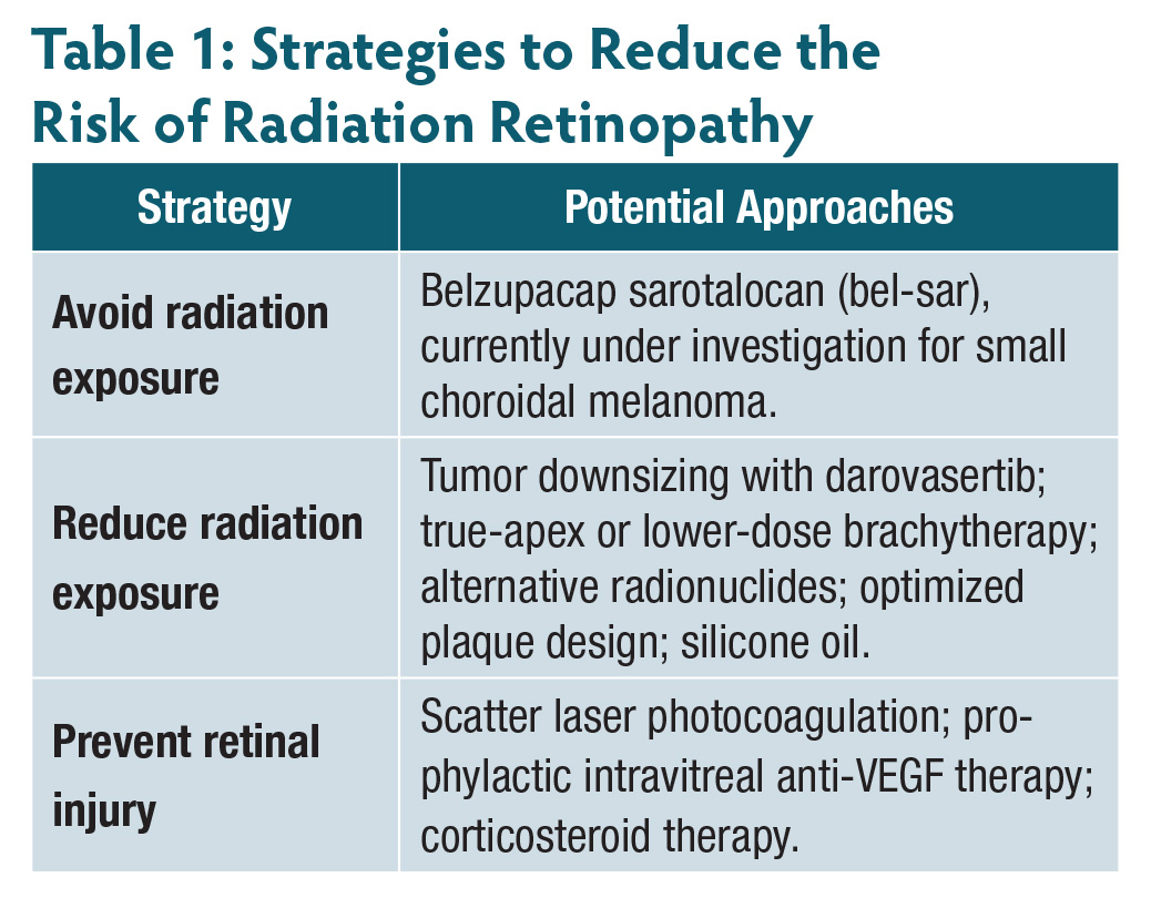
Because effective treatment options are limited, several strategies have been developed to reduce the risk of radiation retinopathy (Table 1). These approaches may be implemented at the time of tumor treatment or during follow-up.
Avoiding Radiation: Nonradiation Treatment Options
For small choroidal melanomas measuring up to 4 mm in height, transpupillary thermotherapy (TTT) was developed as an option that avoids the need for episcleral plaque brachytherapy.3 This method uses an 810-nm infrared laser beam to heat the tumor and induce necrosis. Initial local tumor control has been reported in 94% of cases.3 However, because of concerns regarding delayed recurrence, TTT has largely been abandoned as primary therapy.4
Another nonradiation treatment modality for small choroidal melanoma is belzupacap sarotalocan, or bel-sar (AU-011; Aura Biosciences), which is currently being investigated in the phase 3 randomized, sham-controlled CoMpass trial.5 Bel-sar is a nonreplicating virus-like drug conjugate composed of a photoactive dye linked to a tumor-targeting virus-like particle. The drug is delivered into the suprachoroidal space, allowing the virus-like particle to bind selectively to melanoma cells. When activated with a 689-nm near-infrared diode laser, bel-sar induces reactive oxygen species–mediated tumor necrosis and a secondary proimmunogenic response.
Radiation Dose Reduction
The Collaborative Ocular Melanoma Study (COMS) protocol established an apical dose of 85 Gy as the standard for choroidal melanoma. However, for tumors measuring less than 5.0 mm in height, the prescription point was set at a minimum treatment height of 5.0 mm rather than at the actual tumor apex. In contrast, Singh et al prescribed 85 Gy to the true tumor apex in small choroidal melanomas, achieving excellent local tumor control (99.3%) while reducing radiation exposure to adjacent ocular structures and lowering the risk of radiation retinopathy.6
Recent studies have shown that lower doses, including 65 Gy, may also achieve local tumor control.7 An ongoing investigation is evaluating the efficacy of an apical dose of 63 Gy.8
Alternative Radionuclides and Plaque Designs
The use of different radionuclides can influence radiation dose and scatter. A binuclide radioactive plaque composed of ruthenium 106 (106Ru) and iodine 125 (125I), or a plaque composed entirely of 106Ru, may help spare healthy tissue because of the steep dose gradient produced by the short-range beta decay of 106Ru, particularly for tumors measuring less than 5.0 mm in height.9,10 Similarly, the emission of lower-energy radiation makes palladium 103 a favorable radionuclide for minimizing the risk of radiation retinopathy.11 In addition to custom partial loading of COMS plaques,12 newer plaque designs, such as the EP917 plaque (Eye Physics LLC), may result in lower radiation doses to the macula.13
Reducing Tumor Size
Reducing tumor size before episcleral plaque brachytherapy may decrease radiation exposure and the subsequent risk of radiation retinopathy. Although no drug is currently approved by the US Food and Drug Administration for this purpose, darovasertib (IDE196; Ideaya Biosciences), an oral small-molecule inhibitor of protein kinase C, is being investigated in the randomized phase 3 OptimUM-10 trial involving patients with primary nonmetastatic uveal melanoma. This first-in-class drug targets downstream signaling pathways activated by mutations in the GNAQ and GNA11 genes. One study cohort is evaluating darovasertib before plaque brachytherapy, and another is evaluating the drug before enucleation. The study is expected to be completed in 2031.14
Internal Shielding With Silicone Oil
The use of 1000-centistoke silicone oil at the time of plaque brachytherapy has been shown to attenuate radiation exposure to adjacent ocular tissues, including the macula.15,16 This approach generally involves vitrectomy with silicone oil endotamponade at the time of plaque implantation, followed by silicone oil removal at the time of plaque explantation. The associated risks are those inherent to vitrectomy, including cataract progression, retinal tears, and, rarely, rhegmatogenous retinal detachment.
Laser Photocoagulation
Although scatter laser photocoagulation has been used to slow or halt the progression of radiation retinopathy and maculopathy, it has also been evaluated as a preventive treatment. In patients considered to be at high risk of radiation retinopathy, particularly those with posterior choroidal melanoma, prophylactic scatter laser photocoagulation was associated with a lower rate of radiation retinopathy and less vision loss among patients who developed the condition.11
Intravitreal Anti-VEGF Agents and Corticosteroids
Anti-VEGF agents are a mainstay of treatment for macular edema secondary to radiation retinopathy.17,18 However, their duration of effect appears to be shorter than when they are used to treat diabetic macular edema.19,20 This difference underscores the importance of preventing radiation retinopathy when possible.
In a retrospective comparative study, prophylactic intravitreal injections of bevacizumab (Avastin; Genentech) administered every 4 months after plaque brachytherapy were associated with lower rates of radiation retinopathy and macular edema.21 Shields et al subsequently reported 4-year findings from the same cohort after 2 years of bevacizumab injections administered every 4 months. The expanded analysis included 1,131 treated eyes compared with a historical control group of 117 eyes. Eyes in the bevacizumab group had a lower rate of macular edema at 36 months than eyes in the control group (44% vs 54%; P=.01). Visual acuity outcomes were also better in the bevacizumab group at all evaluated time points, including at 48 months (P<.001).22
Corticosteroids have also been investigated as a prophylactic strategy to reduce radiation-induced retinal injury following plaque brachytherapy. In a randomized controlled trial, the rate and severity of radiation maculopathy among 108 patients who received preventive periocular triamcinolone (Kenalog-40; Bristol Myers Squibb) were compared with those among 55 patients who did not receive corticosteroid treatment. The steroid was administered at the time of plaque placement and again at 4 and 8 months. At 18 months, the rate of moderate visual loss, defined as a loss of 3 or more lines, was significantly lower in the treatment group than in the control group (31% vs 48%; P=.039).23
In another study, a single 0.7-mg intravitreal dexamethasone implant (Ozurdex; AbbVie) was administered to 13 patients with radiation maculopathy. Median central subfield thickness decreased by 120 µm at 1 month (P<.01). However, after 4 months, all patients demonstrated progressive retinal thickening and decreased visual acuity (P<.01).24
The ongoing DRCR Retina Network Protocol AL study is evaluating prophylactic quarterly intravitreal faricimab 6 mg (Vabysmo; Genentech) vs a fluocinolone acetonide 0.19 mg intravitreal implant (Iluvien; ANI Pharmaceuticals) administered every 2 years, with both treatment groups compared with observation for the prevention of radiation retinopathy. This is the first randomized controlled trial to compare these prophylactic treatment strategies.25
Summary
Preventing radiation retinopathy requires a multimodal approach that begins with treatment planning and radiation dose optimization and extends to adjunctive strategies, including prophylactic intravitreal therapy. As new pharmacologic agents, radiation techniques, and molecular therapies are investigated, opportunities to reduce vision-threatening complications following plaque brachytherapy may continue to expand. RP


References
1. Gündüz K, Shields CL, Shields JA, Cater J, Freire JE, Brady LW. Radiation complications and tumor control after plaque radiotherapy of choroidal melanoma with macular involvement. Am J Ophthalmol. 1999;127(5):579-589. doi:10.1016/s0002-9394(98)00445-0
2. Aziz HA, Singh N, Bena J, Wilkinson A, Singh AD. Vision loss following episcleral brachytherapy for uveal melanoma: development of a vision prognostication tool. JAMA Ophthalmol. 2016;134(6):615-620. doi:10.1001/jamaophthalmol.2016.0104
3. Shields CL, Shields JA. Transpupillary thermotherapy for choroidal melanoma. Curr Opin Ophthalmol. 1999;10(3):197-203. doi:10.1097/00055735-199906000-00008
4. Singh AD, Kivelä T, Seregard S, Robertson D, Bena JF. Primary transpupillary thermotherapy of “small” choroidal melanoma: is it safe? Br J Ophthalmol. 2008;92(6):727-728. doi:10.1136/bjo.2007.133249
5. A phase 3 randomized, masked, controlled trial to evaluate efficacy and safety of belzupacap sarotalocan (AU-011) treatment compared to sham control in subjects with primary indeterminate lesions or small choroidal melanoma (CoMpass). Clinicaltrials.gov identifier: NCT06007690. Updated July 31, 2026. Accessed August 5, 2026. https://clinicaltrials.gov/study/NCT06007690
6. Yupari RJ, Bena J, Wilkinson A, Suh J, Singh A. Small choroidal melanoma: outcomes following apical height dose brachytherapy. Br J Ophthalmol. 2021;105(8):1161-1165. doi:10.1136/bjophthalmol-2020-316873
7. Bouhout S, Zhang Z, Suh JH, Singh AD. Low-dose brachytherapy for choroidal melanoma: a preliminary report. Ophthalmol Retina. Published online May 30, 2026. doi:10.1016/j.oret.2026.05.012
8. Kheir WJ, Stinnett SS, Meltsner S, et al. Preliminary results of uveal melanoma treated with iodine-125 plaques: analysis of disease control and visual outcomes with 63 Gy to the target volume. Adv Radiat Oncol. 2022;7(2):100869. doi:10.1016/j.adro.2021.100869
9. Jabbarli L, Fiorentzis M, Rating P, et al. Functional outcome after brachytherapy with bi-nuclide (Ru-106/Iodine-125) plaques in large uveal melanomas. Radiat Oncol. 2025;20(1):22. doi:10.1186/s13014-024-02576-6
10. Wilkinson DA, Kolar M, Fleming PA, Singh AD. Dosimetric comparison of 106Ru and 125I plaques for treatment of shallow ( Br J Radiol. 2008;81(970):784-789. doi:10.1259/bjr/76813976
11. Finger PT, Chin KJ, Duvall G; Palladium-103 for Choroidal Melanoma Study Group. Palladium-103 ophthalmic plaque radiation therapy for choroidal melanoma: 400 treated patients. Ophthalmology. 2009;116(4):790-796.e1. doi:10.1016/j.ophtha.2008.12.027
12. Medek S, Correa ZM, Huth B, Takiar V. Early treatment and toxicity outcomes for small posterior uveal melanomas treated using custom loaded eye plaques. Front Med (Lausanne). 2023;10:1055141. doi:10.3389/fmed.2023.1055141
13. Marwaha G, Wilkinson A, Bena J, Macklis R, Singh AD. Dosimetric benefit of a new ophthalmic radiation plaque. Int J Radiat Oncol Biol Phys. 2012;84(5):1226-1230. doi:10.1016/j.ijrobp.2012.01.084
14. Neoadjuvant darovasertib in primary uveal melanoma. Clinicaltrials.gov identifier: NCT07015190. Updated May 6, 2026. Accessed August 5, 2026. https://clinicaltrials.gov/study/NCT07015190
15. Oliver SC, Leu MY, DeMarco JJ, Chow PE, Lee SP, McCannel TA. Attenuation of iodine 125 radiation with vitreous substitutes in the treatment of uveal melanoma. Arch Ophthalmol. 2010;128(7):888-893. doi:10.1001/archophthalmol.2010.117
16. Ahuja Y, Kapoor KG, Thomson RM, et al. The effects of intraocular silicone oil placement prior to iodine 125 brachytherapy for uveal melanoma: a clinical case series. Eye (Lond). 2012;26(11):1487-1489. doi:10.1038/eye.2012.158
17. Finger PT, Chin KJ, Semenova EA. Intravitreal anti-VEGF therapy for macular radiation retinopathy: a 10-year study. Eur J Ophthalmol. 2016;26(1):60-66. doi:10.5301/ejo.5000670
18. Seibel I, Vollhardt D, Riechardt AI, et al. Influence of ranibizumab versus laser photocoagulation on radiation retinopathy (RadiRet)—a prospective randomized controlled trial. Graefes Arch Clin Exp Ophthalmol. 2020;258(4):869-878. doi:10.1007/s00417-020-04618-7
19. Murray TG, Latiff A, Villegas VM, Gold AS. Aflibercept for Radiation Maculopathy Study: a prospective, randomized clinical study. Ophthalmol Retina. 2019;3(7):561-566. doi:10.1016/j.oret.2019.02.009
20. Yu HJ, Fuller D, Anand R, et al. Two-year results for ranibizumab for radiation retinopathy (RRR): a randomized, prospective trial. Graefes Arch Clin Exp Ophthalmol. 2022;260(1):47-54. doi:10.1007/s00417-021-05281-2
21. Shah SU, Shields CL, Bianciotto CG, et al. Intravitreal bevacizumab at 4-month intervals for prevention of macular edema after plaque radiotherapy of uveal melanoma. Ophthalmology. 2014;121(1):269-275. doi:10.1016/j.ophtha.2013.08.039
22. Shields CL, Dalvin LA, Chang M, et al. Visual outcome at 4 years following plaque radiotherapy and prophylactic intravitreal bevacizumab (every 4 months for 2 years) for uveal melanoma: comparison with nonrandomized historical control individuals. JAMA Ophthalmol. 2020;138(2):136-146. doi:10.1001/jamaophthalmol.2019.5132
23. Horgan N, Shields CL, Mashayekhi A, et al. Periocular triamcinolone for prevention of macular edema after plaque radiotherapy of uveal melanoma: a randomized controlled trial. Ophthalmology. 2009;116(7):1383-1390. doi:10.1016/j.ophtha.2009.01.051
24. Frizziero L, Parrozzani R, Trainiti S, et al. Intravitreal dexamethasone implant in radiation-induced macular oedema. Br J Ophthalmol. 2017;101(12):1699-1703. doi:10.1136/bjophthalmol-2017-310220
25. Intravitreal faricimab injections or fluocinolone acetonide (0.19 mg) intravitreal implants vs observation for prevention of VA loss due to radiation retinopathy (Protocol AL). Clinicaltrials.gov identifier: NCT05844982. Updated July 16, 2026. Accessed August 5, 2026. https://clinicaltrials.gov/study/NCT05844982